


The adaptive immune system relies on its ability to distinguish "self" from "non-self." This distinction is made possible by the Major Histocompatibility Complex (MHC), a group of genes (encoded by HLA genes in humans) that produce specialized surface proteins. These proteins act as molecular "display cases," presenting small fragments of antigens to T cells via their T cell receptors (TCRs). Understanding the structural and functional differences between MHC Class I and Class II is fundamental to grasping how the body orchestrates cellular and humoral immunity.
The Fundamentals of Antigen Presentation
Antigen presentation is the process by which cells capture, process, and display peptide fragments on their surface to alert the immune system. Depending on where a pathogen is located within a cell, the immune system uses one of two distinct pathways: the endogenous pathway or the exogenous pathway.
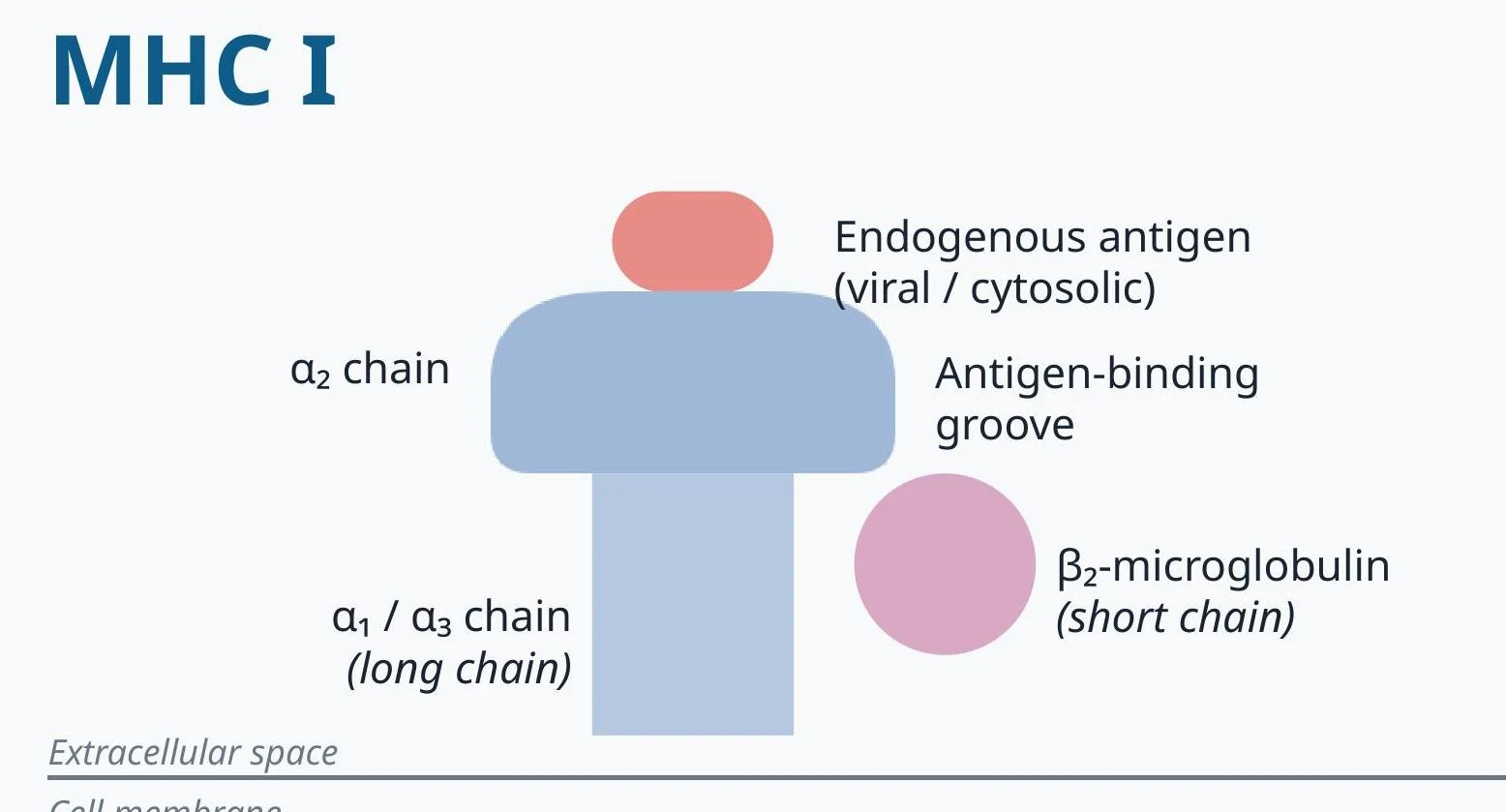
Endogenous Antigens and MHC I
Endogenous antigens are proteins produced within the cytosol of a cell. These typically include normal self-proteins, but also include viral proteins produced during an infection or mutated proteins from a tumor cell.
When these proteins are synthesized, they are broken down into peptides. These peptides are then transported into the endoplasmic reticulum (RER) via a specialized protein complex called TAP (transporter associated with antigen processing). Once inside the RER, the peptides are loaded onto MHC I molecules.
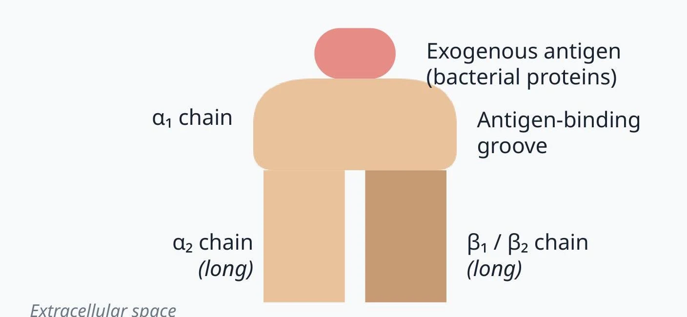
Exogenous Antigens and MHC II
Exogenous antigens are proteins that originate from outside the cell, such as bacteria or extracellular toxins. These are brought into the cell through processes like phagocytosis or endocytosis. Once inside, the antigens are contained within an acidified endosome where they are degraded into peptides. These peptides are then loaded onto MHC II molecules.
Keep reading for free.
Create an account to finish this reading and make it part of your study routine.
Continue reading freeAlready have an account? Log in
Updated