MHC Molecules and Antigen Presentation in the Immune System
Explains the fundamental process of antigen presentation, detailing the roles of MHC Class I and Class II molecules in alerting T cells to threats, and the mechanisms of T cell recognition.
The immune system's ability to distinguish between "self" and "non-self" is one of biology's most sophisticated achievements. Central to this ability is the process of antigen presentation, a mechanism where cells display fragments of proteins (antigens) on their surface to alert T cells of potential threats. Without this presentation, T cells—the orchestrators of adaptive immunity—would remain "blind" to the pathogens invading our cells.
The Fundamentals of Antigen Presentation
Antigen presentation is the process by which an antigen-presenting cell (APC) internalizes an antigen, processes it biochemically into smaller peptides, and displays these peptides on its surface using specialized proteins called Major Histocompatibility Complex (MHC) molecules.
This process is not a single step but a highly regulated pathway involving different cellular compartments. The goal is to ensure that the right type of T cell is activated at the right time: either to kill an infected cell directly or to coordinate a broader immune response via chemical signaling.
This diagram illustrates the process of antigen presentation by MHC class II molecules to CD4+ T cells, a key step in the adaptive immune response. It shows the internalization of an antigen, its processing into peptides within the lysosome, loading onto MHC class II molecules, and finally the presentation of the MHC-peptide complex on the cell surface for recognition by a T cell.
MHC Class I: The Internal Surveillance System
MHC Class I molecules are found on almost all nucleated cells in the body. Their primary role is to report on the internal health of the cell. If a cell is infected by a virus or has become cancerous, it will produce "endogenous" proteins that do not belong there.
The pathway for MHC Class I follows these steps:
Synthesis: Proteins are synthesized within the cell.
Processing: If the proteins are foreign (like viral proteins) or improperly folded self-proteins, they are broken down into peptides.
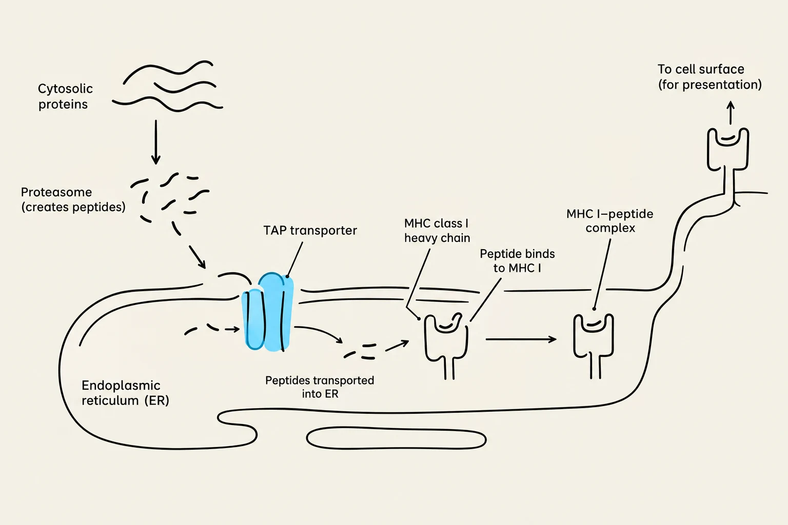
Loading in the ER: These peptides are transported into the Endoplasmic Reticulum (ER).
Transport: Specialized proteins known as TAP-1 and TAP-2 (Transporters associated with Antigen Processing) facilitate the movement of these peptides into the ER to meet the MHC Class I molecules.
Presentation: Once the peptide is bound, the MHC Class I-peptide complex is transported to the cell surface.
Exam Tip: Remember that MHC Class I is "universal" for nucleated cells and presents to CD8+ Cytotoxic T cells. If a cell displays a foreign peptide on MHC I, it is essentially saying, "I am infected; kill me."
MHC Class II: The External Scout System
Unlike Class I, MHC Class II molecules are restricted to professional antigen-presenting cells (APCs), such as macrophages, B cells, and dendritic cells. Their job is to monitor the extracellular environment for "exogenous" antigens—pathogens that have been swallowed by the cell.
The MHC Class II pathway is distinct and involves several specialized components:
The Invariant Chain (Ii): When MHC Class II molecules are synthesized in the ER, they are not allowed to bind peptides immediately. Instead, they are complexed with the Invariant chain (Ii). This chain serves two purposes: it prevents the MHC II molecule from binding endogenous peptides intended for MHC I, and it helps guide the molecule toward the correct cellular compartment.
Endosomal and Lysosomal Compartments: The MHC II molecule travels from the ER to specialized acidic compartments called endosomes or lysosomes.
Peptide Loading: In these compartments, the Invariant chain is removed, and the MHC II molecule encounters peptides derived from phagocytosed (swallowed) antigens.
Presentation: The final MHC Class II-peptide complex is then moved to the cell surface.
Diagram illustrating the process of antigen presentation via MHC class I molecules, involving cytosolic proteins, proteasomes, TAP transporters, and the endoplasmic reticulum.
T Cell Recognition: CD4+ vs. CD8+
The specificity of the immune response relies on the interaction between the T-cell receptor (TCR) and the MHC-peptide complex. T cells are categorized based on the "co-receptor" they use to bind to the MHC molecule:
Feature
MHC Class I
MHC Class II
Target T Cell
CD8+ (Cytotoxic T cells)
CD4+ (Helper T cells)
Antigen Source
Endogenous (inside the cell)
Exogenous (outside the cell)
Cell Distribution
All nucleated cells
Professional APCs
Immune Role
Killing infected/abnormal cells
Coordinating the immune response
T-Cell Receptors (TCRs)
T cells recognize antigens through their TCRs, which are heterodimers (usually α and β chains). Unlike antibodies (immunoglobulins), TCRs exist only on the cell surface and do not undergo somatic mutation to increase diversity. Instead, their massive variety is generated through V-J recombination during development. To signal the cell after an antigen is recognized, the TCR is associated with the CD3 complex and zeta (ζ) chains.
Antigenic Specificity: Haptens and Carriers
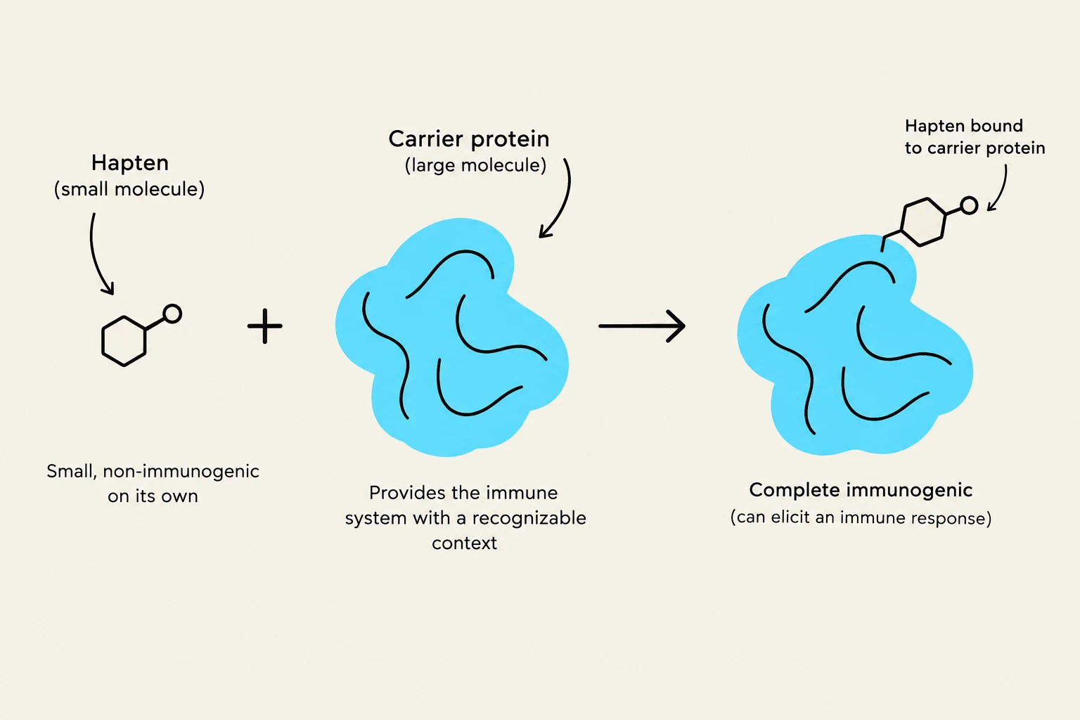
To understand how the immune system recognizes specific shapes, we must look at the relationship between antigens and antibodies. Sometimes, a molecule is too small to trigger an immune response on its own; these are called haptens.
A hapten (such as penicillin or certain toxins) is not immunogenic by itself. However, if the hapten attaches to a larger, more complex molecule known as an immunogenic carrier, it becomes part of a new, larger antigen. The immune system then recognizes the hapten as a novel antigenic determinant of that carrier. This principle is vital in studying antigen-antibody reactions and understanding how allergies or vaccine responses are triggered.
This diagram illustrates how a hapten, a small molecule that is not immunogenic on its own, can become immunogenic when bound to a carrier protein. The carrier protein provides a recognizable context for the immune system, allowing it to elicit an immune response.