This article explains precipitation reactions in immunology, where soluble antigens and antibodies form insoluble complexes. It details three key methods: Rocket Immunoelectrophoresis for antigen quantification, Immunofixation Electrophoresis for detecting antibodies and antigens (especially in multiple myeloma diagnos
In immunology, the interaction between an antigen (AGN) and an antibody (ABY) can result in various visible or measurable phenomena. While agglutination involves the clumping of particulate antigens, precipitation refers to the formation of insoluble complexes when soluble antigens and antibodies meet in the right proportions. To make these interactions easier to detect, scientists often use gel electrophoresis, a technique that combines an electric current with diffusion to achieve faster and sharper results.
By utilizing electrophoresis, we can separate molecules based on their size and charge, allowing for the precise identification and quantification of specific antigen-antibody complexes. This article explores three primary methods: Rocket Immunoelectrophoresis, Immunofixation Electrophoresis, and the Western Blot.
Rocket Immunoelectrophoresis
Rocket immunoelectrophoresis is a specialized technique used primarily to quantitate the immunoglobulins (antibodies) present in body fluids. In this method, the antibody is already known and is distributed throughout a gel, while the unknown antigen is placed into a well within that gel.
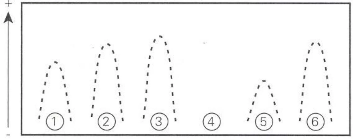
When an electric current is applied, electrophoresis facilitates the migration of the antigens toward the antibodies. As they meet, they form antigen-antibody complexes that precipitate out of the solution. This creates a characteristic "rocket-shaped" pattern in the gel.
A diagram illustrating separation based on charge or size, with numbered bands representing different components.
The key to this method is its quantitative nature. The height of the "rocket" is directly proportional to the amount of antigen present in the sample. By constructing a standard curve, laboratory professionals can determine the exact concentration of the antigen in a patient's sample.
Exam Tip: Remember that in Rocket Immunoelectrophoresis, the antibody is known and the antigen is unknown. The height of the peak tells you the concentration of the antigen.
Immunofixation Electrophoresis (IFE)
Immunofixation electrophoresis (IFE) is a highly sensitive method used to detect both antibodies and antigens. Unlike the rocket method, IFE is a two-stage process typically performed on agarose or cellulose acetate.
Separation: The test material (containing unknown antigens) undergoes gel electrophoresis to separate the different proteins.
Fixation: Specific antiserum is added to the gel. The antibodies in the antiserum diffuse into the gel to react with the separated antigens, forming complexes. The gel is then washed to remove any non-precipitated proteins, and the remaining precipitated proteins are stained for examination.
Clinical Application: Multiple Myeloma Diagnosis
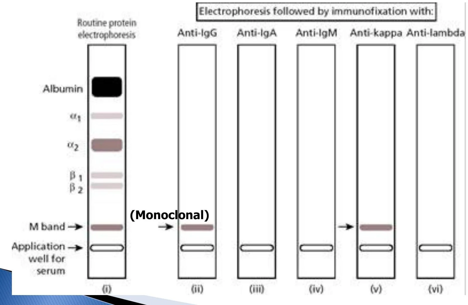
IFE is a critical diagnostic tool for identifying multiple myeloma, a type of cancer where malignant plasma cells produce an excess of a single type of antibody. By electrophoresing a patient's serum or urine, clinicians can identify "monoclonal" bands (often called M-bands) that indicate the presence of these abnormal proteins.
This diagram illustrates protein electrophoresis and immunofixation, showing the separation of proteins like albumin and the identification of a monoclonal band.
This image displays the results of an immunofixation electrophoresis (IFE) test, likely showing protein bands separated by electrical charge and then identified by specific antibodies. The labels EP, G, A, M, K, and lambda likely represent different proteins or immunoglobulin chains being analyzed.
Western Blot
The Western Blot is an adaptation of immunofixation electrophoresis used to detect specific antibodies against complex antigens. It is most famously used in the diagnostic protocols for infectious diseases such as HIV, Lyme disease, and syphilis.
The Procedure
The Western Blot involves several precise steps to ensure the antigens are transferred and detected correctly:
Electrophoresis: Known antigens are separated on a gel.
Blotting: The gel is placed in a "sandwich" with nitrocellulose paper. An electric field is applied to pull the protein bands out of the gel and absorb them onto the paper.
Incubation: The patient's serum is added to the paper, and the mixture is incubated and rinsed.
Detection: The paper is stained, and the presence of specific bands is examined.
Diagram illustrating the process of preparing an agarose gel, including mixing reagents, boiling, cooling, pouring into a mold, and forming wells with a comb.
Diagram illustrating the steps of a Western blot assay, showing antigen-antibody binding, enzyme-labeled antibody incubation, and substrate reaction leading to color development.
HIV Detection and Diagnostic Algorithms
In the context of HIV, Western Blotting is often used as a confirmatory test following a reactive initial screen. Modern diagnostic algorithms typically follow a tiered approach:
Initial Screen: An FDA-approved HIV-1/2 antigen/antibody combination immunoassay.
Differentiation: If the screen is reactive, an antibody differentiation immunoassay is used.
Confirmatory/Alternative: If results are indeterminate, a nucleic acid test (NAT) may be utilized.
When performing a Western Blot for HIV, clinicians look for specific recombinant HIV antigens. A positive result is typically reported if at least two of the following three bands are present:
p24
gp 41
gp 120/gp 160
Troubleshooting Electrophoresis
Electrophoretic techniques are highly sensitive to experimental conditions. Errors can lead to inaccurate results or failed runs. Common errors include:
Applying the electric current in the wrong direction.
Using an incorrect pH or allowing the separation to run for too long (or too short) a time.
Using a current that is too weak or too strong.
Improper ratios of antigen to antibody, which can prevent the formation of visible complexes.
Keep reading for free.
Create an account to finish this reading and make it part of your study routine.