Explores the diverse roles of membrane proteins, including receptors, metabolic enzymes, and transport proteins. Discusses passive and active transport mechanisms, the physics of electrochemical gradients, and provides a clinical connection to hemoglobin mutations and sickle cell anemia.
Biological membranes are not merely passive barriers; they are dynamic environments regulated by a diverse array of specialized proteins. These proteins allow cells to communicate with their surroundings, perform metabolic reactions, and precisely control the movement of molecules across the lipid bilayer. Understanding how these proteins function is fundamental to grasping how cells maintain homeostasis and respond to environmental changes.
Major Classes of Membrane Proteins
Membrane proteins are categorized based on their specific biological roles. While they all reside within or span the membrane, their functions vary significantly.
Membrane Receptor Proteins
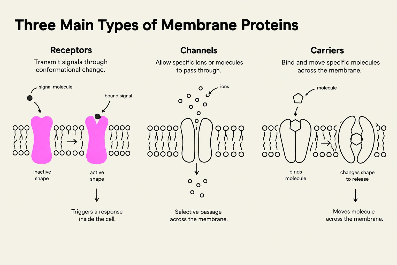
Receptors are responsible for signal transduction. A classic example is the G Protein-Coupled Receptor (GPCR). These proteins transmit extracellular signals (such as hormones or neurotransmitters) into the cell without the physical movement of the signaling molecule itself. When a ligand binds to the extracellular domain, it triggers a conformational change in the protein's structure, which subsequently activates internal signaling pathways within the cell.
Membrane-Bound Metabolic Enzymes
Some proteins serve as catalysts for biochemical reactions. These enzymes are often embedded in membranes, such as the inner mitochondrial membrane, where they facilitate critical redox reactions that drive processes like ATP production.
Membrane Transport Proteins
Transport proteins are essential for moving polar or charged molecules—which cannot easily pass through the hydrophobic core of the lipid bilayer—across the membrane. These are further divided into passive and active transporters based on their energy requirements.
Diagram illustrating the three main types of membrane proteins: receptors, channels, and carriers, and their respective functions in cell signaling and transport.
The Physics of Transport: Electrochemical Gradients
To understand how molecules move, we must define the "driving force" behind their motion. For any molecule, movement is governed by its electrochemical gradient. This gradient consists of two distinct components:
The Chemical Component: The difference in concentration of the molecule across the membrane. Molecules naturally tend to move from an area of high concentration to low concentration.
The Electrical Component: The difference in electrical charge across the membrane (the membrane potential). This component only affects charged ions.
The total free energy change (ΔG) associated with moving an ion across a membrane can be calculated using the following relationship:
ΔG=RTln(C1C2)+zFΔψ
In this equation, C1 is the initial concentration, C2 is the final concentration, R is the gas constant, T is the temperature, z is the charge of the ion, F is Faraday's constant, and Δψ is the electrical potential. For uncharged molecules, the electrical component becomes zero because z=0.
Exam Tip: When calculating ΔG, remember that a negative value indicates movement "down" the gradient (a favorable process), while a positive value indicates movement "against" the gradient (an unfavorable process requiring energy).
Passive Transport: Channels and Carriers
Passive transport occurs when molecules move "down" their electrochemical gradient, meaning the process is spontaneous and requires no metabolic energy (such as ATP).
Channels
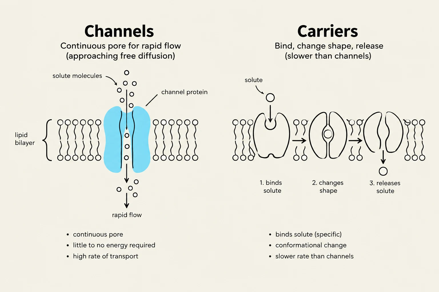
Channels are highly selective pores that allow specific ions or molecules to pass through the membrane at speeds approaching free diffusion—the rate at which molecules move through a solution without obstruction.
A vital example is the aquaporin, a channel specifically designed for water transport. Aquaporins must be highly selective to allow water to pass while preventing "proton jumping" (the leakage of $H^+$ ions). They achieve this through specific selectivity filters and the orientation of their internal dipoles, which forces water molecules to reorient as they pass, breaking the continuous chain of hydrogen bonds required for proton movement.
Carriers
Unlike channels, carriers bind specifically to a substrate, which induces a conformational change to move the molecule to the other side. Because of this required binding step, carriers are slower than channels and are subject to saturation; once all binding sites are occupied, the transport rate reaches a maximum capacity (Vmax). This process is often referred to as facilitated diffusion.
Diagram illustrating the difference between channel proteins and carrier proteins in facilitated diffusion across a cell membrane.
Active Transport: Moving Against the Gradient
When a cell needs to accumulate a substance against its concentration gradient, it must use active transport. Active transport can only be performed by carriers.
Primary Active Transport
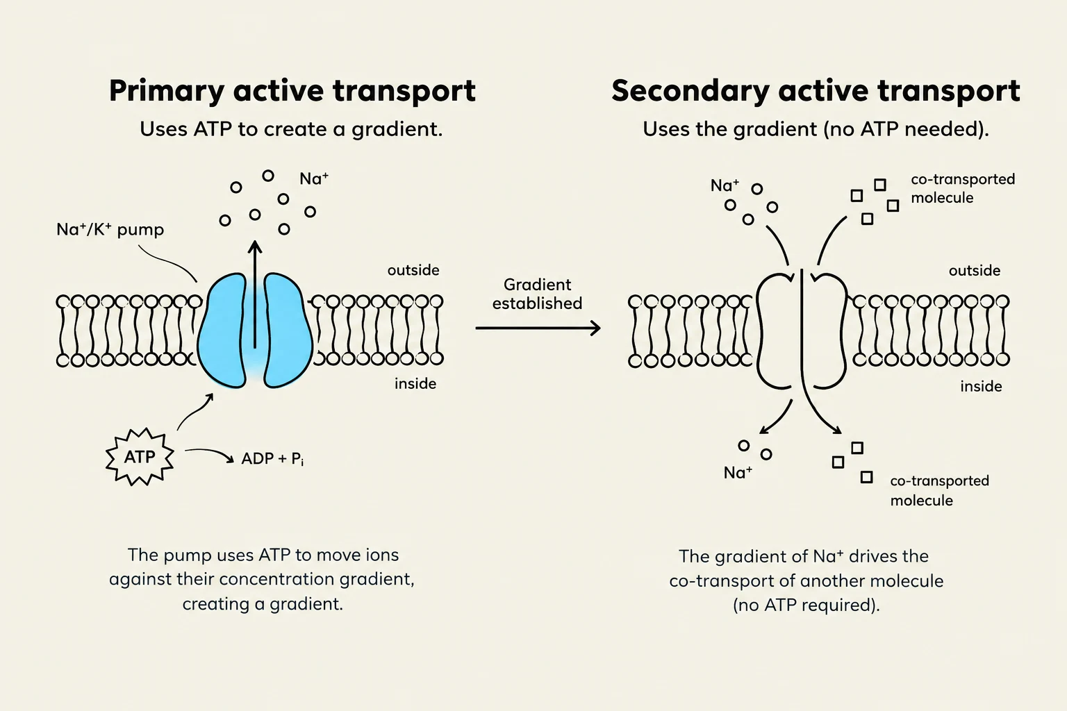
This mechanism directly utilizes metabolic energy, most commonly from ATP hydrolysis, to "pump" a substrate across the membrane.
Secondary Active Transport
Secondary active transport does not use ATP directly. Instead, it uses the energy stored in an existing electrochemical gradient (usually created by a primary active transporter) to drive the movement of a second molecule. This is achieved through two modes:
Symport: The carrier moves two different molecules in the same direction.
Antiport: The carrier moves two different molecules in opposite directions.
Diagrams illustrating primary and secondary active transport across a cell membrane, showing the roles of ATP and ion gradients.
Clinical Connection: Hemoglobin Mutations
The importance of protein structure and conformational change is clearly illustrated in hemoglobin, the protein responsible for oxygen transport. Mutations in the genes encoding hemoglobin can lead to various diseases, such as sickle cell anemia.
In sickle cell anemia, a single point mutation replaces a hydrophilic glutamate with a hydrophobic residue at position six of the β-chain. This creates a hydrophobic patch on the exterior of the protein. Because hydrophobic groups tend to aggregate to avoid water, deoxygenated hemoglobin molecules stick together, forming long, insoluble fibers. These fibers distort the red blood cell into a "sickle" shape, which can block capillaries and cause severe pain and organ damage.
Keep reading for free.
Create an account to finish this reading and make it part of your study routine.