Explains the fundamental concept of membrane potential in all cells, focusing on how excitable cells like neurons and muscle fibers utilize and rapidly alter this potential for communication and function. Covers ion distribution, resting membrane potential, electrochemical gradients, the sodium-potassium pump, and chan
In the study of physiology, few concepts are as fundamental as the electrical charge of a cell. Every living cell in the human body maintains a charge difference across its plasma membrane. This phenomenon, known as membrane potential, acts much like a biological battery, storing potential energy that can be harnessed to perform work. While all cells possess a membrane potential, "excitable cells"—specifically those in nervous and muscle tissues—have the unique ability to deliberately and rapidly alter this potential to facilitate communication and movement.
The Foundation: Membrane Potential and Ion Distribution
To understand how a cell functions, we must first look at the environment surrounding it. A cell is divided into two distinct compartments: the intracellular fluid (the fluid inside the cell) and the extracellular fluid (the fluid outside the cell). Dissolved within these fluids are various ions, which are atoms or molecules that carry an electrical charge.
The distribution of these ions is not equal. The specific arrangement of these charged particles creates the membrane potential. Key players in this distribution include:
Potassium ($K^+$): The primary ion found in higher concentrations inside the cell.
Sodium (Na+): Found in much higher concentrations outside the cell.
Other Ions: Calcium (Ca2+), Chlorine ($Cl^-$), and various large proteins also contribute to the charge. Notably, at physiological pH, these proteins carry a negative charge and are too large to pass through the membrane.
This unequal distribution results in a typical "resting" state where the inside of the cell is negatively charged relative to the outside.
Diagram illustrating the difference in ion concentrations between extracellular and intracellular fluid across a cell membrane, highlighting the roles of sodium (Na+) and potassium (K+) ions, and negative proteins.
The Resting Membrane Potential (RMP)
When a cell is not actively sending signals, it is said to be at its resting membrane potential (RMP). This is the baseline electrical state of the cell. While different cell types have different RMP values (for example, a neuron might be at −70 mV while a skeletal muscle cell is at −90 mV), the principle remains the same: the interior is more negative than the exterior.
The RMP is maintained by the constant interplay of two forces acting on ions, collectively known as the electrochemical gradient:
Chemical (Concentration) Gradient: This force drives ions to move from areas of high concentration to areas of low concentration via facilitated diffusion through protein channels.
Electrical Gradient: This force drives ions toward opposite charges (opposites attract).
The Tug-of-War: Potassium vs. Sodium
The movement of ions is often a battle between these two gradients. For potassium, the chemical gradient pushes the ion out of the cell (where concentration is lower), while the electrical gradient tries to pull the positive ion in (toward the negative interior). In most cases, the chemical gradient is much stronger, resulting in a net movement of $K^+$ out of the cell.
Conversely, for sodium, both the chemical and electrical gradients work in the same direction, driving the ion into the cell "like gang busters".
Exam Tip: Do not confuse permeability with the volume of ion flow. Permeability refers to the possibility of movement based on the number of available leak channels (protein channels that are always open). The cell membrane is significantly more permeable to potassium than to sodium because there are more potassium leak channels available, even though the electrochemical gradient for sodium is much stronger.
Maintaining the Battery: The Sodium-Potassium Pump
If ions were allowed to move freely through leak channels indefinitely, they would eventually reach equilibrium, meaning there would be no net movement and no charge difference. A cell at equilibrium is a dead cell.
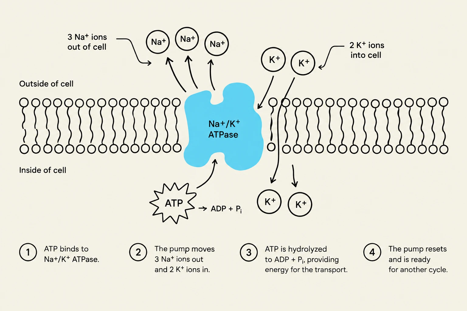
To prevent this, the cell uses a specialized protein called the sodium-potassium ATPase (or pump). This is a form of active transport that requires energy in the form of ATP to move ions against their concentration gradients. The pump works via an antiport mechanism, moving ions in opposite directions:
It pumps three sodium (Na+) ions out of the cell.
It pumps two potassium ($K^+$) ions in to the cell.
This process is incredibly energy-intensive; it is estimated that approximately 90% of the ATP produced in your body is used just to run these pumps.
Diagram illustrating the mechanism of the Na+/K+-ATPase pump, which transports sodium ions out of the cell and potassium ions into the cell, utilizing ATP hydrolysis for energy.
Excitable Cells: Changing the Potential
While all cells maintain an RMP, excitable cells (neurons and muscle fibers) are specialized to change their membrane potential rapidly in response to a stimulus. This change can be electrical, chemical, or physical.
When these cells change their potential, they produce two types of electrical events:
Graded Potentials: Localized changes that only affect a specific part of the membrane.
Action Potentials: Changes that conduct across the entire membrane rapidly.
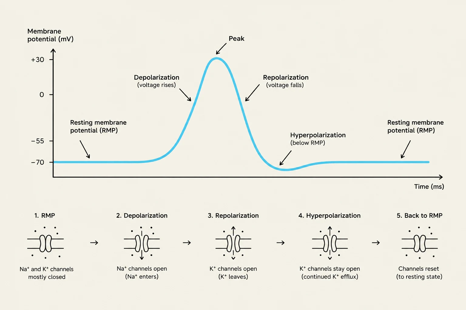
Depolarization, Repolarization, and Hyperpolarization
The direction of the membrane potential change depends on which ion channels open and which way the ions flow:
Depolarization: When the membrane potential becomes more positive (moving closer to zero or above). This typically happens when sodium or calcium channels open, allowing positive ions to rush into the cell.
Repolarization: The process of the membrane potential returning from a depolarized state back down to the resting membrane potential.
Hyperpolarization: When the membrane potential becomes more negative than the RMP. This can happen if potassium channels open (allowing positive ions to leave) or if chlorine ($Cl^-$) channels open (allowing negative ions to enter).
This diagram illustrates the changes in membrane potential during an action potential, showing depolarization, repolarization, and hyperpolarization, along with the corresponding states of ion channels.
Application in Neurons and Muscle
In a neuron, the dendrites and soma primarily contain chemical-gated ion channels, which respond to neurotransmitters to create graded potentials. The axon, however, is specialized with voltage-gated ion channels that allow for the rapid conduction of action potentials.
In muscle cells, the process is similar. The motor end plate (the area where a nerve meets the muscle) utilizes chemical-gated channels to initiate a signal, while the rest of the muscle fiber membrane is covered in voltage-gated channels to ensure the entire cell contracts in unison.
Summary Table: Ion Movement and Potential Change
Ion
Direction of Movement
Effect on Membrane Potential
Sodium (Na+)
Inward
Depolarization (More Positive)
Potassium ($K^+$)
Outward
Hyperpolarization (More Negative)
Calcium (Ca2+)
Inward
Depolarization (More Positive)
Chlorine ($Cl^-$)
Inward
Hyperpolarization (More Negative)
Keep reading for free.
Create an account to finish this reading and make it part of your study routine.