Explores the dynamic nature of biological membranes, focusing on how cells synthesize, assemble, and maintain the crucial asymmetry of phospholipid distribution within the lipid bilayer. Covers processes in the endoplasmic reticulum and Golgi apparatus, and the importance of this asymmetry for cellular functions like s
Biological membranes are not merely static barriers; they are dynamic, two-dimensional fluids that serve as the fundamental architecture of life. While the lipid bilayer is characterized by its fluidity—allowing proteins and lipids to move laterally within the plane of the membrane—it is also highly organized. One of the most striking features of these membranes is their asymmetry: the two leaflets (monolayers) of the bilayer contain distinct sets of molecules, creating unique chemical identities for the "inside" and "outside" faces of the cell. Understanding how cells build these membranes and maintain this precise distribution is essential to understanding cellular signaling, trafficking, and survival.
Membrane Assembly in the Endoplasmic Reticulum
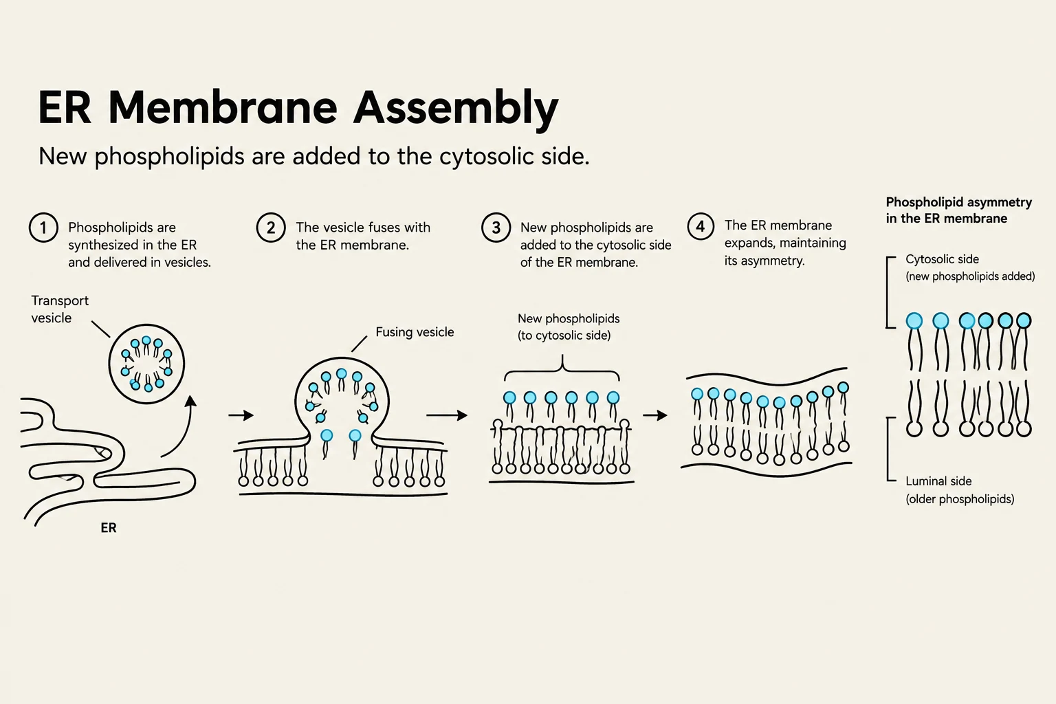
The lifecycle of a membrane begins in the endoplasmic reticulum (ER), the cell's primary factory for lipid synthesis. In the ER, enzymes bound to the cytosolic surface of the membrane use free fatty acids as substrates to synthesize new phospholipids.
Because these enzymes are located exclusively on the cytosolic side, they can only add new lipids to the cytosolic monolayer. If left unchecked, this would cause the membrane to grow unevenly, making one side much larger than the other. To prevent this, the ER employs a specialized transporter protein called scramblase. Scramblases randomly transfer phospholipids from the cytosolic monolayer to the noncytosolic monolayer, ensuring that the ER membrane grows symmetrically.
Diagram illustrating the process of ER membrane assembly, where new phospholipids are added to the cytosolic side, expanding the membrane while maintaining its asymmetry.
Study Tip: Remember the distinction between the ER and the Golgi. In the ER, the goal is symmetry (via scramblases), whereas in the Golgi, the goal is asymmetry (via flippases).
Establishing Asymmetry in the Golgi Apparatus
As membranes move from the ER to the Golgi apparatus, they undergo a profound transformation. While the ER membrane is relatively uniform, the Golgi membrane is highly asymmetric. This is achieved by a different class of transporters known as flippases.
Unlike scramblases, which move lipids randomly and do not require energy, flippases are highly selective and use the energy from ATP hydrolysis to move specific phospholipids from the noncytosolic side to the cytosolic side. For example, flippases actively move phosphatidylserine and phosphatidylethanolamine to the cytosolic monolayer. This leaves other lipids, such as phosphatidylcholine and sphingomyelin, concentrated in the noncytosolic monolayer. This selective "flipping" establishes the chemical distinction between the interior of an organelle and the exterior environment.
Preservation of Membrane Orientation
A critical requirement for cellular function is that this asymmetry is preserved during transport. When a vesicle buds from one organelle and fuses with another (such as the plasma membrane), the membrane maintains its orientation: the cytosolic monolayer always remains facing the cytosol, and the noncytosolic monolayer remains facing the exterior or the lumen of an organelle.
This preservation is vital for several reasons:
Protein Function: Membrane proteins must be oriented correctly to interact with the outside world or to transport molecules into the cell.
Glycolipids: These lipids have sugar groups attached to their hydrophilic heads. Because these sugars are added in the Golgi on the noncytosolic side and there are no flippases to move them, they remain "trapped" in that monolayer. Consequently, when these lipids reach the plasma membrane, their sugars are correctly positioned to face the extracellular environment, forming the glycocalyx (a protective sugar coating).
Diagram illustrating the function of flippase in the Golgi apparatus, which uses ATP to move lipids between the outer and inner leaflets of the cell membrane.
Internalization: Endocytosis and Sorting
Cells must also bring materials from the outside into the interior. This is achieved through various forms of endocytosis:
Phagocytosis: The "eating" of large particles (like bacteria) using cellular extensions called pseudopods.
Pinocytosis: The non-specific uptake of extracellular fluid via vesicles.
Receptor-Mediated Endocytosis: A highly efficient process where specific molecules (like LDL/cholesterol, iron, or Vitamin B12) bind to surface receptors, concentrating the cargo and increasing internalization efficiency by over 1000-fold.
Once internalized, materials enter the endosome compartment. The endosomal lumen is kept acidic (pH≈5–6) by an ATP-driven $H^+$ pump. This acidity is a critical "sorting signal"; it causes many receptors to release their cargo, allowing the receptors to be recycled back to the plasma membrane while the cargo moves forward.
Intracellular Digestion and Autophagy
The final destination for many endocytosed materials is the lysosome, the cell's primary digestive organelle. Lysosomes contain approximately 40 different types of hydrolytic enzymes (including proteases and lipases) that are optimally active at an acidic pH≈5. To prevent the lysosome from digesting itself, its membrane proteins are heavily glycosylated, creating a protective sugar shield.
Beyond bringing in external materials, the cell also uses this system to clean itself through autophagy. In this process, the cell encloses obsolete or damaged organelles within a double membrane to form an autophagosome, which then fuses with a lysosome for degradation. This allows the cell to recycle essential components like amino acids during periods of starvation.
Keep reading for free.
Create an account to finish this reading and make it part of your study routine.