Explores the fundamental physics of image quality in medical imaging, including spatial and contrast resolution, signal-to-noise ratio (SNR), and contrast-to-noise ratio (CNR). It also covers signal propagation, attenuation, and the evaluation of diagnostic accuracy using Receiver Operating Characteristic (ROC) analysi
In clinical medicine, no single imaging modality is perfect for every diagnostic task. Choosing the right tool—whether it be X-ray, MRI, CT, or ultrasound—requires a deep understanding of how different energies interact with human tissue and how we measure the accuracy of the resulting images. This article explores the fundamental physics of image quality, the challenges of signal loss, and the statistical methods used to evaluate diagnostic performance.
Fundamentals of Image Quality
To determine if an image is "good" enough for a diagnosis, clinicians look at several interrelated physical properties.
Spatial and Contrast Resolution
Spatial resolution is the ability to distinguish between two closely spaced objects. If two structures, such as the liver and the stomach, have high spatial resolution, their boundaries will appear clear and distinct. If resolution is poor, these structures may appear to blend together.
Contrast resolution refers to how well different tissues can be distinguished from one another. Even if an image has high resolution (fine detail), it may be useless if the contrast between two tissues is too low to tell them apart.
SNR and CNR: The Role of Noise
Every imaging system records some level of noise, which is any unwanted signal not related to the actual anatomy being measured. To quantify the quality of the signal, we use two ratios:
Signal-to-Noise Ratio (SNR): This compares the desired signal to the unwanted noise.
SNR=NoiseDesired signal
A higher SNR indicates a cleaner, more reliable image.
Contrast-to-Noise Ratio (CNR): This describes how well two different tissues can be distinguished specifically in the presence of noise. High SNR does not automatically guarantee high CNR; you must have both a strong signal and a significant difference in signal intensity between tissues to achieve high CNR.
Exam Tip: Remember that improving one factor (like resolution) does not automatically improve others (like SNR). They must work together to produce a diagnostically useful image.
This illustration explains the concepts of spatial resolution, signal-to-noise ratio (SNR), and contrast-to-noise ratio (CNR) in imaging. It shows how higher spatial resolution can sometimes reduce CNR if not managed properly.
Signal Propagation and Attenuation
Imaging modalities use different forms of energy, often described by their position on the electromagnetic spectrum. These waves are characterized by their wavelength (λ) and frequency (f). In the case of ultrasound, we deal with mechanical pressure waves rather than electromagnetic waves.
The Concept of Attenuation
As a signal travels through a medium (like human tissue), it loses strength. This total propagation loss is called attenuation. Attenuation is caused by four primary interactions:
Absorption: The energy is taken up by the tissue and converted into heat.
Reflection: The signal hits a boundary and some energy returns toward the source.
Refraction: The wave changes direction when entering a medium with a different propagation velocity.
Scattering: The signal is dispersed in different directions.
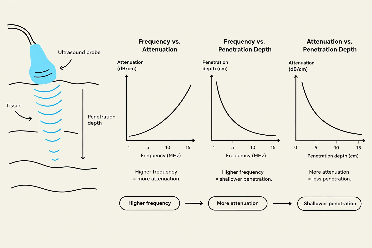
In ultrasound, attenuation is highly dependent on frequency. High-frequency waves provide better resolution but suffer from high attenuation (low penetration), whereas low-frequency waves provide better penetration but lower resolution.
This diagram illustrates the relationship between ultrasound frequency, attenuation, and penetration depth in tissue. Higher frequencies lead to greater attenuation and shallower penetration, while lower frequencies penetrate deeper but are attenuated more.
Evaluating Accuracy: ROC Analysis
Once an image is captured, we must evaluate how well the diagnostic test or imaging system distinguishes between healthy and diseased states. This is done using Receiver Operating Characteristic (ROC) analysis.
The Four Outcomes
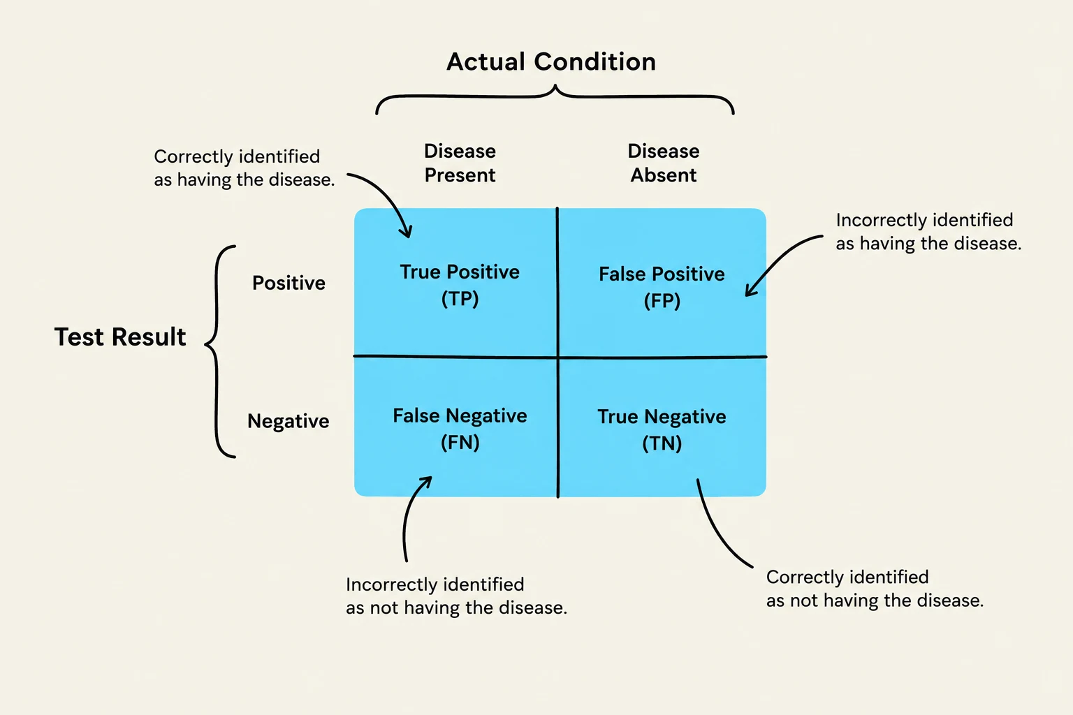
To perform this analysis, we categorize every test result into one of four outcomes:
True Positive (TP): The condition is present, and the test correctly identifies it.
False Positive (FP): The condition is absent, but the test incorrectly indicates it is present.
True Negative (TN): The condition is absent, and the test correctly identifies it.
False Negative (FN): The condition is present, but the test incorrectly indicates it is absent.
Key Performance Metrics
We quantify the performance of a modality using three mathematical metrics:
1. Accuracy
The proportion of total correct diagnoses (both true positives and true negatives):
Accuracy=Total diagnosesCorrect diagnoses×100%
2. Sensitivity
The ability of a test to correctly identify those who actually have the condition. High sensitivity is crucial for minimizing False Negatives:
Sensitivity=TP+FNTP×100%
3. Specificity
The ability of a test to correctly identify those who do not have the condition. High specificity is crucial for minimizing False Positives:
Specificity=TN+FPTN×100%
Clinical Note: In many medical contexts, a False Negative is considered more dangerous than a False Positive. A false positive (e.g., a suspicious mammogram) usually leads to more testing, whereas a false negative (e.g., missing a cancer diagnosis) can lead to untreated, life-threatening illness.
A confusion matrix illustrating the outcomes of a diagnostic test, showing true positives, true negatives, false positives, and false negatives in relation to the actual condition.
Clinical Application and Decision Making
ROC analysis is not just a theoretical exercise; it guides real-world clinical and economic decisions. For example, a hospital with a limited budget must decide whether to invest in an MRI, a CT scanner, or nuclear medicine equipment based on the specific patient populations they serve and the required diagnostic accuracy for those populations.
Similarly, the choice of modality depends on the specific goal. Ultrasound is often selected for pregnancy imaging because it provides useful diagnostic information without the use of ionizing radiation. By understanding the trade-offs between resolution, attenuation, and statistical accuracy, clinicians can select the most effective tool for the patient's needs.
Keep reading for free.
Create an account to finish this reading and make it part of your study routine.