Explains how the lymphatic system collects excess interstitial fluid, forming lymph, and the physiological mechanisms, including muscular activity and pressure gradients, that transport it back to the circulatory system. It also touches upon the immune functions of the lymphatic system.
The lymphatic system serves as a vital secondary circulatory network, acting as both a drainage system for excess fluids and a primary theater for immune defense. Unlike the cardiovascular system, which utilizes the heart as a centralized pump to circulate blood, the lymphatic system is a low-pressure, one-way system that relies on various physiological mechanisms to move fluid from the tissues back toward the heart. Understanding how lymph is formed and how it is propelled is essential for grasping how the body maintains fluid homeostasis and protects itself from pathogens.
The Genesis of Lymph: Interstitial Fluid and Osmotic Balance
At the microscopic level, blood capillaries constantly exchange fluids with the surrounding tissues. While most fluid is reabsorbed into the venous end of capillaries, a portion remains in the spaces between cells, known as interstitial fluid. This excess interstitial fluid is collected by lymphatic capillaries, at which point it is officially termed lymph.
The rate of lymph formation is heavily influenced by the balance of pressures within the capillary beds. A critical factor is osmotic pressure, which is primarily maintained by plasma proteins, most notably albumin. Albumin acts like a "molecular sponge," exerting an osmotic pull that draws fluid from the interstitial space back into the blood capillaries.
When there is a decrease in osmotic pressure—often due to a deficiency in albumin levels—the blood's ability to retain fluid diminishes. This results in more fluid remaining in the interstitial space, which subsequently increases the production of lymph.
This diagram illustrates how albumin levels in blood plasma affect osmotic pressure and fluid balance. Higher albumin levels increase osmotic pressure, drawing water into capillaries. Conversely, lower albumin levels decrease osmotic pressure, leading to less water being pulled into capillaries and potentially causing the accumulation of interstitial fluid.
Exam Tip: If a question asks what triggers an increase in lymph formation, look for "decrease in osmotic pressure" or "reduction in albumin."
Clinical Implications: Obstruction and Lymphedema
Because the lymphatic system is responsible for the lymphatic return (the process of bringing fluid back to the central circulation), any interruption in this pathway can lead to significant clinical issues.
A common example occurs in oncology patients. During surgery for breast cancer, axillary lymph nodes (located in the armpit) are often removed to prevent the spread of cancer cells. However, removing these nodes can cause a disorder in the return of lymph from the arm, leading to a condition known as lymphedema. This results in chronic, heavy swelling of the limb.
Patients suffering from lymphedema face a heightened risk of infection because the affected area is "poorly protected" due to the compromised lymphatic drainage. Consequently, medical professionals advise extreme caution with these limbs:
Avoid taking blood pressure readings on the affected arm.
Avoid injections in the swollen area.
Avoid minor skin traumas, such as manicures, which could introduce pathogens that the lymphatic system cannot effectively clear.
Mechanisms of Lymph Transport
Since the lymphatic system lacks a central pump, it utilizes a combination of active and passive mechanisms to move lymph through the body toward the thoracic duct and eventually the heart.
1. Muscular Activity and Skeletal Contraction
The most significant driver of lymph movement is muscular activity. When we move—such as when walking—the contraction of skeletal muscles physically compresses the lymphatic vessels, "milking" the lymph forward through the system.
2. Smooth Muscle Contraction
The walls of the larger lymphatic vessels contain smooth muscle. The rhythmic contraction of this smooth muscle provides an intrinsic pumping action that helps propel the fluid along the vessel.
3. The Respiratory Pump
The act of breathing also assists in transport. As the diaphragm moves during inhalation and exhalation, it creates pressure changes within the thoracic and abdominal cavities. These pressure fluctuations act as a "pump," helping to draw lymph and blood toward the heart.
4. Lymphatic Valves
To ensure that lymph moves in only one direction, lymphatic vessels are equipped with internal valves. These valves allow for unidirectional flow and prevent the backflow of lymph due to gravity or pressure changes.
Diagram illustrating the function of valves within a lymphatic vessel, showing how they open to allow lymph flow forward and close to prevent backflow.
The Immune Connection: Chemical Signaling
The lymphatic system is not just a drainage network; it is the primary highway for immune cells. When the body encounters a threat, various chemical signals are released to coordinate a defense.
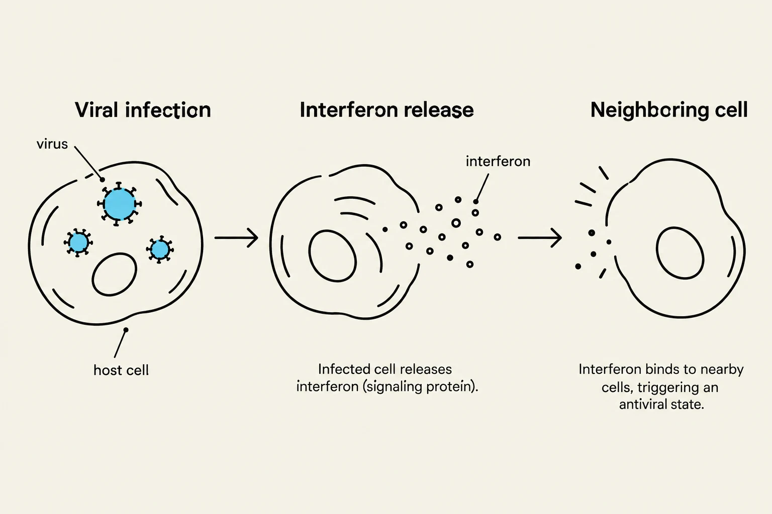
Interferon: This is a specific chemical released by cells that have been invaded by a viral infection. The interferon acts as a warning signal, alerting neighboring cells to activate their antiviral defenses.
Cytokines and Chemokines: While interferon is specific to viruses, other chemicals like cytokines and chemokines are released by tissues in response to general injury or the presence of parasites. These chemicals act as signals to attract immune cells to the site of the injury or infection.
Through these mechanisms, the lymphatic system ensures that fluid balance is maintained and that the body's immune response is rapidly and effectively deployed.
This diagram illustrates the process of viral infection and the subsequent release of interferon, a signaling protein, by an infected cell. The interferon then binds to nearby cells, triggering an antiviral state.
Keep reading for free.
Create an account to finish this reading and make it part of your study routine.