An overview of the human immune system, detailing the innate and adaptive immune responses, the inflammatory process, fever, and the roles of B and T lymphocytes, antigen presentation, and MHC molecules.
The human immune system is a sophisticated, multi-layered defense network designed to protect the body from internal and external threats, including pathogens like bacteria, viruses, and fungi, as well as cancerous cells. This defense is not a single entity but a highly integrated coordination between the innate immune system (the rapid, non-specific first and second lines of defense) and the adaptive immune system (the slower, highly specific third line of defense).
The Inflammatory Response: The Second Line of Defense
When pathogens bypass surface barriers like the skin or mucous membranes, the innate immune system initiates an inflammatory response. Inflammation is a non-specific response to cellular injury caused by trauma, toxins, or pathogens. It serves to attract immune cells to the site of damage, clear away debris, and facilitate healing.
Stage 1: Release of Mediators and Cardinal Signs
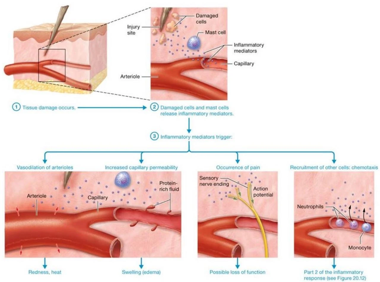
The process begins when damaged cells and local mast cells release various inflammatory mediators. These chemicals include histamine, serotonin, cytokines, bradykinin, prostaglandins, and leukotrienes.
These mediators trigger the "cardinal signs" of inflammation:
Vasodilation: Mediators like histamine and bradykinin relax smooth muscle in arterioles, increasing blood flow to the area (hyperemia). This results in redness and heat.
Increased Capillary Permeability: The "leakiness" of local capillaries increases, allowing protein-rich fluid to escape into tissue spaces, which causes swelling (edema).
Pain: Bradykinin and prostaglandins trigger sensory neurons, signaling the body that tissue damage is occurring.
This diagram illustrates the process of inflammation, starting with tissue damage that triggers the release of inflammatory mediators. These mediators cause vasodilation, increased capillary permeability, pain, and the recruitment of immune cells like neutrophils and monocytes to the injury site.
Stage 2: Phagocyte Response
Once the area is prepared, white blood cells (leukocytes) are recruited to the site via chemotaxis (movement in response to chemical signals).
Macrophages: Local macrophages act as "first responders," activating within minutes to ingest pathogens and debris.
Neutrophils: These are the most numerous granulocytes and are highly effective at destroying bacteria. They undergo margination (adhering to capillary walls) and diapedesis (squeezing through endothelial gaps) to enter the tissue.
Monocytes: These circulate in the blood and migrate to the tissue to differentiate into additional macrophages.
The accumulation of dead leukocytes, dead tissue, and fluid at the site forms a whitish mixture known as pus.
Exam Tip: If a clinical lab report shows leukocytosis (an elevated white blood cell count), it is a general indicator that inflammation or infection is occurring somewhere in the body.
Fever: The Systemic Innate Response
While inflammation is a local response, fever is a systemic innate response. Fever is initiated when chemicals called pyrogens are released from damaged cells or certain bacteria. These pyrogens act on the hypothalamus, which functions as the body's thermostat, causing it to reset the body's temperature set point to a higher value.
Adaptive Immunity: B and T Lymphocytes
The third line of defense is adaptive (acquired) immunity. Unlike the innate system, it responds to unique glycoprotein markers called antigens. It is characterized by its ability to "remember" an antigen, allowing for a much faster and more efficient response upon re-exposure.
B Cells and Humoral Immunity
B lymphocytes (B cells) are responsible for humoral immunity (antibody-mediated immunity). They mature in the bone marrow.
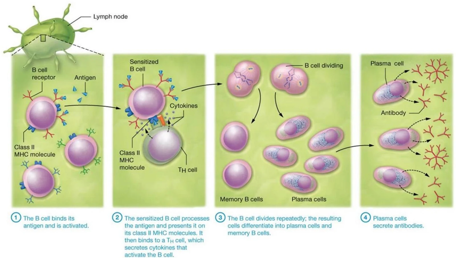
Activation: Through clonal selection, a specific antigen binds to a B cell receptor. To become fully activated, the B cell often presents the antigen on Class II MHC molecules to a Helper T cell.
Differentiation: Once activated, B cells proliferate and differentiate into:
Plasma cells: These secrete antibodies (immunoglobulins).
Memory B cells: These provide long-term, rapid protection during subsequent exposures.
This illustration depicts the process of B cell activation and differentiation, leading to antibody production. It shows how a B cell binds to an antigen, interacts with a T helper cell, divides, and differentiates into plasma cells that secrete antibodies and memory B cells.
T Cells and Cell-Mediated Immunity
T lymphocytes (T cells) are responsible for cell-mediated immunity, which targets intracellular pathogens (like viruses), cancer cells, and foreign transplants. T cells mature in the thymus.
There are three primary types of T cells:
Helper T (TH) cells: These are the "coordinators." They secrete cytokines to stimulate other cells. For example, they use interleukin-3 to stimulate macrophages and interleukin-2 to activate Cytotoxic T cells.
Cytotoxic T (TC) cells: These directly destroy diseased or foreign cells by releasing perforins (which create pores in the target cell membrane) and enzymes that induce apoptosis (programmed cell death).
Memory T cells: These provide long-term immunity without needing the initial co-stimulators.
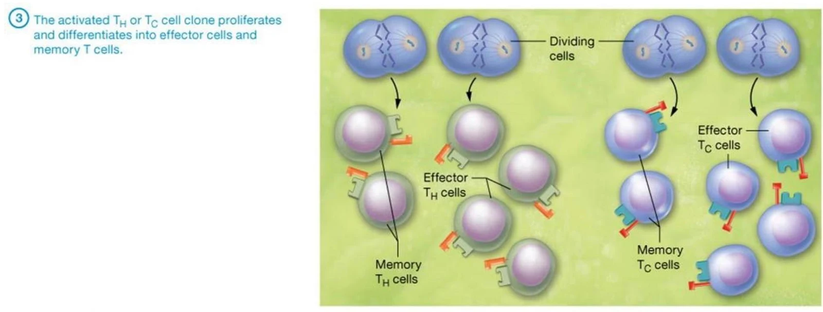
This illustration depicts the proliferation and differentiation of activated T helper (TH) or cytotoxic T (Tc) cells into effector and memory T cells.
Antigen Presentation and MHC Molecules
For T cells to function, they must "see" antigens. However, T cells cannot recognize free-floating antigens; they only recognize antigens that are bound to Major Histocompatibility Complex (MHC) molecules on the surface of other cells.
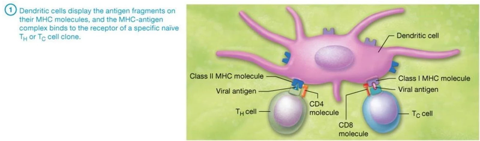
Class I MHC: Found on nearly all nucleated cells. They present endogenous antigens (antigens synthesized inside the cell, such as viral proteins or cancer antigens) to TC cells.
Class II MHC: Found only on Antigen-Presenting Cells (APCs), such as dendritic cells and macrophages. They present exogenous antigens (antigens taken in via phagocytosis) to TH cells.
Dendritic cells are considered the most important APCs because they possess the unique ability of cross-presentation, where they can display ingested antigens on both Class I and Class II MHC molecules to activate both TC and TH cells.
This illustration shows how dendritic cells present antigen fragments on MHC molecules to T cells, initiating an immune response.
Transplantation and Rejection
The immune system uses MHC molecules to distinguish "self" from "non-self." This is the basis for transplant rejection:
Autografts: Tissue from the same individual (no rejection).
Isografts: Tissue between genetically identical individuals (no rejection).
Allografts: Tissue between nonidentical individuals of the same species (high risk of rejection).
Xenografts: Tissue between different species (high risk of rejection).
Rejection occurs when TC cells recognize foreign MHC antigens (encoded by HLA genes) on the transplanted tissue.
Summary of Antibody Functions
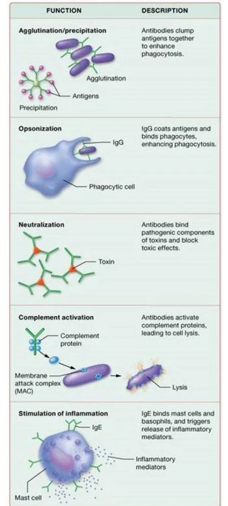
Antibodies (immunoglobulins) are Y-shaped proteins produced by plasma cells that neutralize or destroy antigens.
Class
Primary Functions
IgG
Most abundant in serum; can cross the placenta; functions in opsonization and neutralization
IgA
Found in secretions (saliva, breast milk); functions in agglutination and neutralization
IgM
First antibody secreted; largest; potent agglutinating and precipitating agent
Found on the surface of B cells; aids in B cell activation
This diagram illustrates five functions of antibodies in the immune system: agglutination/precipitation, opsonization, neutralization, complement activation, and stimulation of inflammation.
Keep reading for free.
Create an account to finish this reading and make it part of your study routine.