


Homologous recombination is a fundamental biological process involving the exchange of genetic material between homologous DNA molecules. This mechanism is essential for maintaining genomic integrity through DNA repair and, in eukaryotes, serves as a programmed and vital component of meiotic prophase I. By facilitating the exchange of information between similar DNA sequences, recombination ensures genetic diversity and the accurate segregation of chromosomes.
Initiation: Double-Strand Breaks
The process of homologous recombination is almost always initiated by double-strand breaks (DSBs), where both strands of the DNA duplex are severed. The management of these breaks varies significantly between prokaryotes and eukaryotes.
In bacteria, such as E. coli, the RecBCD pathway is the primary mechanism for managing DSBs. The RecBCD complex acts as both a helicase and a nuclease, processing the broken DNA ends to create the necessary single-stranded segments required for the next steps of the pathway.
In eukaryotes, the initiation is often more controlled, particularly during meiosis. The enzyme Spo11 is responsible for creating programmed DSBs to facilitate recombination.
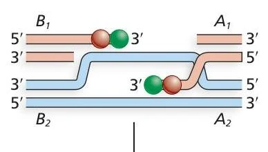
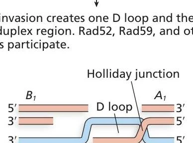
Following the initial break, specialized proteins like Mrx and Exo1 (which are homologs of bacterial helicases and nucleases) trim the DNA to generate long, single-stranded overhangs.
Strand Invasion and D-loop Formation


Keep reading for free.
Create an account to finish this reading and make it part of your study routine.
Continue reading freeAlready have an account? Log in
Updated