The plasma membrane of a cell acts as a sophisticated gatekeeper. While the hydrophobic interior of the lipid bilayer serves as a natural barrier to most hydrophilic molecules—including essential ions, sugars, and amino acids—cells must find ways to move these substances across the membrane to survive. To overcome this barrier, cells utilize specialized membrane transport proteins that facilitate the movement of solutes. These proteins do not all work the same way; some simply allow molecules to flow down their natural gradients, while others act as "pumps" that harness energy to move substances against their concentration gradients.
The Driving Force: Electrochemical Gradients
To understand how transport proteins work, we must first understand the "force" that drives them. For uncharged molecules like glucose, movement is dictated solely by the concentration gradient—the difference in the amount of solute between the inside and outside of the cell. Substances naturally flow "downhill" from high concentration to low concentration via passive diffusion.
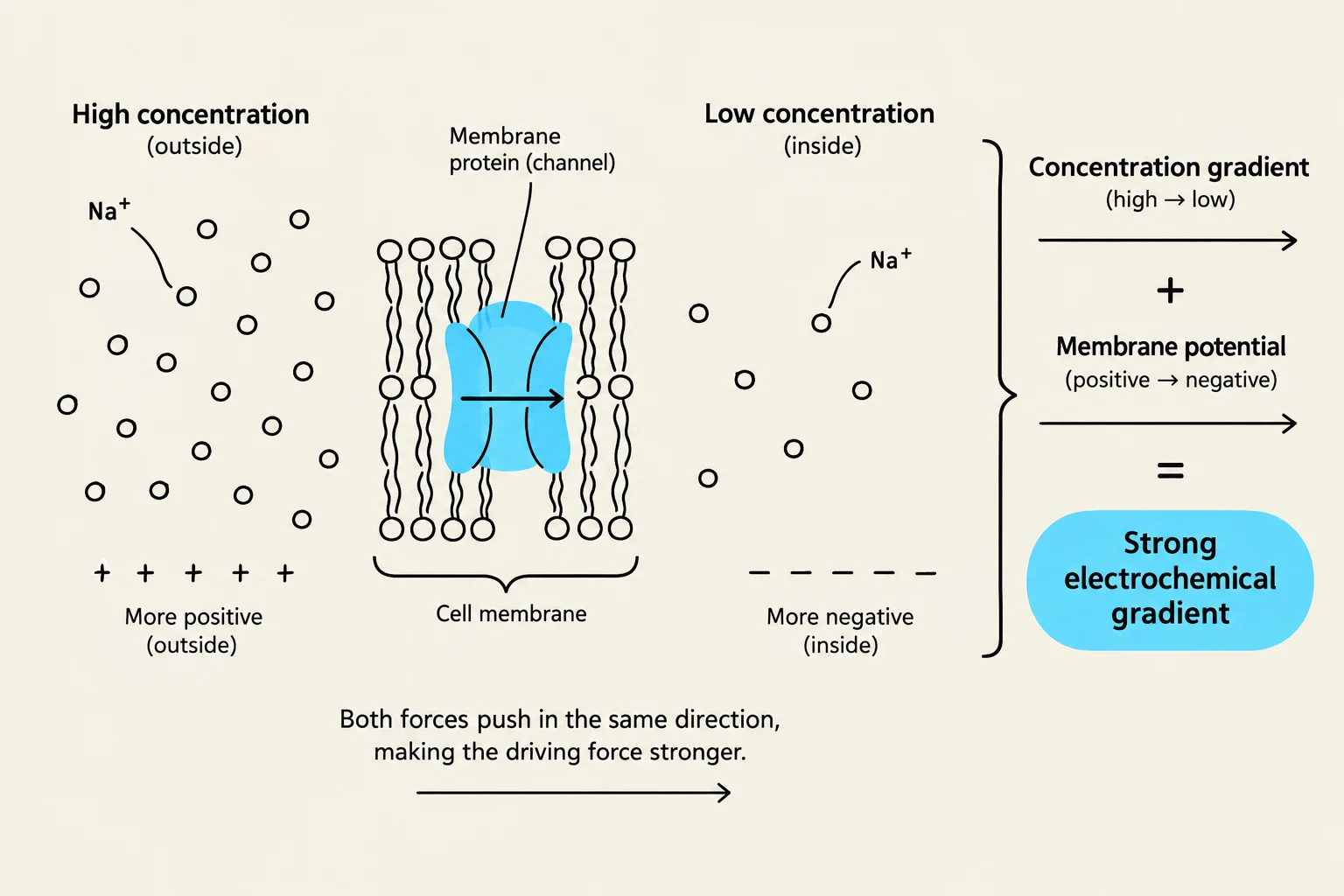
However, for charged particles (ions), the situation is more complex. Because the interior of a cell is typically more negatively charged than the exterior, a voltage difference known as the membrane potential exists across the plasma membrane. The movement of an ion is therefore determined by the sum of two forces: the concentration gradient and the electrical force from the membrane potential. This combined net driving force is called the electrochemical gradient.
For example, Na+ is highly concentrated outside the cell and is also positively charged, meaning both the concentration gradient and the negative interior of the cell pull Na+ inward, creating a very steep electrochemical gradient.
This diagram illustrates the concept of an electrochemical gradient across a cell membrane, showing how the combined forces of concentration difference and membrane potential drive ion movement.
Exam Tip: Remember that for uncharged molecules, the electrochemical gradient is simply the concentration gradient. For ions, you must account for both the concentration difference and the electrical charge of the membrane.
Categorizing Transporters: Uniports, Symports, and Antiports
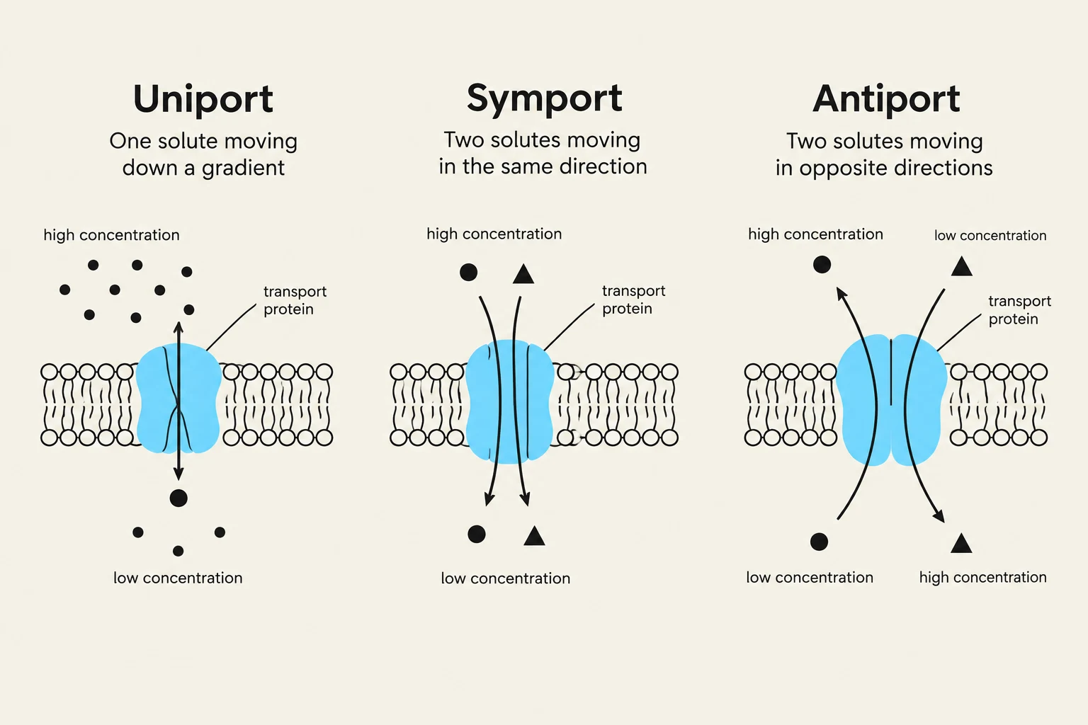
Transporters are categorized based on how many solutes they move and the direction in which they move them.
Uniports
A uniport facilitates the passive transport of a single type of solute down its concentration gradient. A classic example is the glucose transporter found in many mammalian cells. The protein switches between different conformational states—outward-open, inward-open, and an occluded state—allowing glucose to move from an area of high concentration to low concentration. Because uniports move solutes "downhill" without an external energy source, they are not considered pumps.
Symports (Coupled Transport)
Symports are a type of coupled transport where a pump moves two different solutes in the same direction across the membrane. This mechanism allows a cell to move a substance "uphill" (against its gradient) by using the "downhill" energy of another substance.
A vital biological example is the glucose–Na+ symport. The cell uses the steep inward electrochemical gradient of Na+ to drive the active import of glucose into the cell, even when glucose concentrations are already higher inside the cytosol.
Antiports (Coupled Transport)
In antiport mechanisms, the pump moves two solutes in opposite directions. This is often used to regulate the internal environment of the cell. For instance:
The Na+–H+ exchanger uses the influx of Na+ to pump $H^+$ (protons) out of the cell, which is essential for regulating cytosolic pH.
The Ca2+/Na+ exchanger moves Ca2+ out of muscle cells to facilitate muscle relaxation.
Diagrams illustrating Uniport, Symport, and Antiport transport mechanisms across a cell membrane, showing the movement of solutes via transport proteins.
Gradient-Driven Pumps and the Role of ATP
While symports and antiports use existing gradients, they rely on a "master" mechanism to keep those gradients from disappearing: gradient-driven pumps.
In animal cells, the ATP-driven Na+ pump (also known as the Na+–K+ ATPase) is the engine of the cell. It uses energy from ATP hydrolysis to pump Na+ out of the cell and $K^+$ into the cell. This process is vital because Na+ is constantly "leaking" back into the cell through other channels and transporters. Without the Na+ pump constantly working to expel Na+, the electrochemical gradient would vanish, and all other gradient-driven symports and antiports would cease to function.
Spatial Organization in Epithelial Cells
In specialized tissues, such as the gut epithelium, cells use these mechanisms in a highly organized spatial pattern to achieve net transport of nutrients from the environment into the body.
Apical Domain (facing the gut lumen): Symports are located here to pull nutrients like glucose into the cell from the gut.
Basal Domain (facing the bloodstream): Uniports are located here to allow the glucose to exit the cell and enter the blood.
This directional flow is only possible because of tight junctions, which act as a diffusion barrier between the apical and basal domains, preventing the solutes from simply leaking back to where they started.
Summary Table: Transport Mechanisms
Mechanism
Directionality
Energy Source
Example
Uniport
Single solute
Concentration gradient
Glucose transporter
Symport
Two solutes, same direction
Electrochemical gradient
Glucose–Na+ symport
Antiport
Two solutes, opposite direction
Electrochemical gradient
Na+–H+ exchanger
Pump
Against gradient
ATP or Light
Na+–K+ ATPase
Keep reading for free.
Create an account to finish this reading and make it part of your study routine.