Explores how transcription factors bind to specific DNA sequences using structural motifs and interactions with major and minor grooves to regulate gene expression.
Every cell in a multicellular organism contains the exact same genome, yet a neuron looks and functions entirely differently from a hepatocyte. This incredible diversity is not due to the presence or absence of genes, but rather to transcriptional control—the fundamental "on/off" switch that determines which genes are expressed at any given time. By regulating transcription, cells can achieve specialization, allowing them to develop unique structural and molecular features while maintaining a shared genetic blueprint.
The Logic of Transcriptional Control
Gene expression is a multi-layered process, but transcription is the primary level of regulation because it serves as the basis for cell differentiation and memory. While "housekeeping genes" (such as those for glycolysis or ribosomal proteins) are expressed in almost all cells to maintain basic survival, "tissue-specific genes" (like insulin in pancreatic cells) are turned on only when needed.
To control this process, the cell utilizes transcription factors (TFs). These are proteins that bind to specific DNA sequences to either recruit or block RNA polymerase, thereby modulating the rate of transcription. Because the human genome contains over 3 billion base pairs, a transcription factor must be able to find its specific 6–20 bp target site with immense precision.
Reading the Code: Major and Minor Grooves
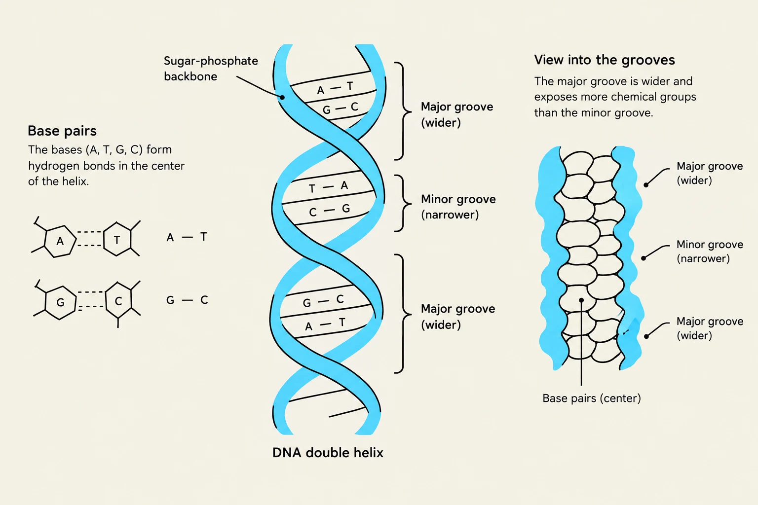
How do proteins "read" a DNA sequence without unwinding the double helix? The answer lies in the physical structure of the DNA molecule itself. The double helix creates two distinct channels: the major groove and the minor groove.
The major groove is wider and exposes a unique pattern of chemical groups for each base pair combination. Specifically, it presents a distinct arrangement of hydrogen-bond donors, hydrogen-bond acceptors, and hydrophobic methyl groups (found on thymine). Because each of the four base pairs (A-T, T-A, G-C, C-G) presents a unique chemical "signature" in the major groove, transcription factors can achieve high sequence-specific recognition by making direct contact with these groups.
The minor groove is narrower and provides less sequence-specific information. However, some proteins utilize the minor groove to recognize the overall shape or flexibility of the DNA, such as identifying AT-rich regions which tend to be narrower and more flexible.
Diagram illustrating the structure of a DNA double helix, including its sugar-phosphate backbone, base pairs (A-T and G-C), and the major and minor grooves.
Structural Motifs of DNA Binding
Transcription factors use various modular structural motifs to achieve their binding goals. While the "recognition element" (often an α-helix) makes specific contacts with the bases, "stabilizing elements" grip the phosphate backbone via electrostatic interactions to hold the protein in place.
Helix-Turn-Helix (HTH)
One of the oldest and most common motifs, especially in bacteria, consists of two α-helices connected by a short turn. The second helix, known as the recognition helix, inserts directly into the major groove to make base-specific contacts. These proteins often bind as dimers to palindromic sequences, where each monomer reads one half of the symmetry.
Homeodomain Motif
The homeodomain is a highly conserved domain of approximately 60 amino acids featuring three α-helices. In this motif, Helix 3 acts as the recognition helix in the major groove, while a flexible N-terminal arm reaches into the minor groove to provide additional specificity. These are famous as "master regulators" of embryonic development; for example, Hox genes use homeodomains to dictate segment identity along the body axis in organisms ranging from flies to humans.
Leucine Zipper Motif
This motif is characterized by two α-helices that contain a leucine residue every seven amino acids. These leucines interlock like a zipper to form a coiled-coil dimer, creating a Y-shaped structure. The "arms" of the Y contain positively charged basic regions that grip the DNA backbone and insert into the major groove.
Zinc Finger Motif
The zinc finger is a small, finger-shaped loop stabilized by a single zinc ion (Zn2+) coordinated by cysteine and histidine residues. These motifs are highly modular and often appear in tandem arrays. For example, a protein with three zinc fingers can recognize a 9 bp sequence, providing massive specificity (49 possible combinations).
Helix-Loop-Helix (HLH) Motif
In the HLH motif, two α-helices are connected by a flexible loop. While the HLH structure itself facilitates dimerization, a separate basic region is responsible for the actual DNA binding. This motif is critical for cell differentiation; for instance, the MyoD protein uses this mechanism to drive the development of muscle cells.
Exam Tip: When comparing motifs, remember that dimerization is a universal theme. Dimerization increases both the specificity of the target site and the regulatory flexibility of the cell, as different combinations of proteins (homodimers vs. heterodimers) can recognize different sets of genes.
Increasing Specificity and Cooperativity
A single transcription factor binding to a 6 bp site might find thousands of accidental matches in a large genome. To solve this, cells use dimerization and cooperative binding.
When two monomers bind together as a dimer, they can recognize a much longer sequence (e.g., 12 bp), which drastically reduces the number of random occurrences in the genome. Furthermore, many TFs exhibit cooperative binding, where the binding of one protein facilitates the binding of another nearby.
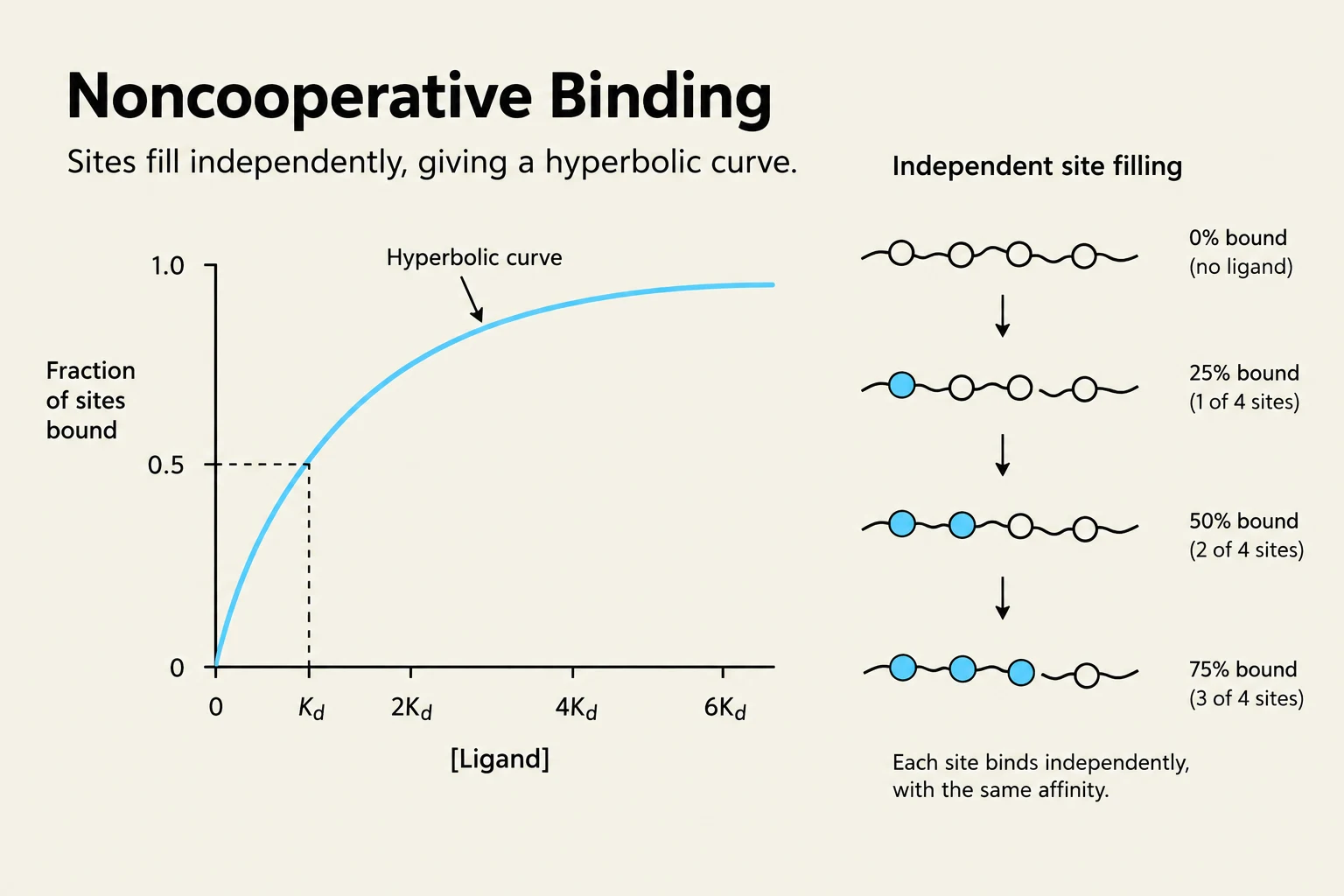
Unlike noncooperative binding, which follows a gradual hyperbolic curve, cooperative binding produces a sigmoidal (S-shaped) curve. This allows for a sharp "all-or-none" transition from an OFF state to an ON state in response to small changes in signal concentration. This sensitivity is vital during embryonic development, where precise, rapid decisions must be made.
This illustration depicts noncooperative binding, where sites fill independently, resulting in a hyperbolic curve. The right side shows a step-by-step process of ligand binding to four sites, illustrating 0%, 25%, 50%, and 75% binding.
The Chromatin Barrier and Pioneer Factors
In eukaryotes, DNA is not always "naked" and accessible; it is wrapped around histone proteins to form nucleosomes. This creates a physical barrier to transcription factors. Most TFs require "nucleosome-free" DNA to bind.
To overcome this, cells use two main strategies:
Chromatin Remodeling: Activators recruit enzymes like Histone Acetyltransferases (HATs) to acetylate histones, which in turn recruits remodelers that slide nucleosomes along the DNA to expose binding sites.
Pioneer Factors: These are special transcription factors that have the unique ability to bind DNA even when it is still wrapped around a nucleosome. Once bound, they partially destabilize the nucleosome and recruit remodeling complexes to open the chromatin for other, more conventional TFs.
By combining these structural motifs, cooperative interactions, and chromatin-modifying tools, the cell achieves the sophisticated level of control necessary for complex life.
Keep reading for free.
Create an account to finish this reading and make it part of your study routine.