This content explains the fundamental mechanisms behind autoimmune disorders, detailing how the immune system can mistakenly target the body's own tissues. It covers the roles of self-reactive cells and antigens, and explores four primary pathways leading to autoimmunity: release of sequestered antigens, molecular mimi
In a healthy immune system, a critical process called "self-tolerance" ensures that the body can distinguish between foreign invaders and its own healthy tissues. During the development of immune cells, the body undergoes a selection process; for instance, in the bone marrow, approximately 90% of B cells that recognize self-antigens are destroyed to prevent them from attacking the body. However, when this regulatory system fails, the immune system begins to target the body's own components.
This breakdown leads to the activation of self-reactive T cells and self-reactive B cells that bind to self-antigens. Instead of protecting the organism, these cells initiate an immune response against healthy cells, leading to chronic inflammation and tissue damage.
The Players: Self-Reactive Cells and Antigens
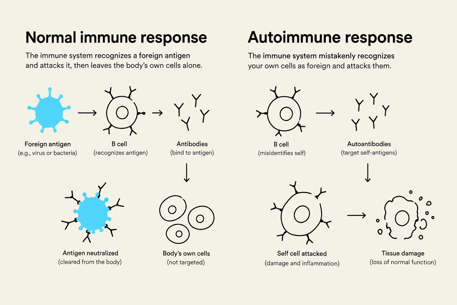
To understand autoimmunity, we must first understand the components involved in a standard immune response. In a normal humoral (antibody-mediated) response, B cells bind to specific antigens via their B cell receptors. Once activated, these B cells differentiate into plasma cells, which secrete antibodies, and memory B cells, which provide long-term protection.
In an autoimmune context, the "antigen" is not a pathogen like a virus or bacteria, but a self-antigen—a normal molecule belonging to the body's own cells. When self-reactive T cells or B cells encounter these self-antigens, they treat them as threats.
This diagram illustrates the difference between a normal immune response, where the body attacks foreign antigens like viruses or bacteria, and an autoimmune response, where the immune system mistakenly attacks the body's own cells, leading to tissue damage.
Exam Tip: Remember that the primary difference between an infection and an autoimmune disorder is the nature of the antigen. In infection, the antigen is foreign; in autoimmunity, the antigen is a "self-antigen" that the immune system has failed to recognize as "self".
Primary Mechanisms of Autoimmunity
Researchers have identified four distinct pathways through which the immune system loses its ability to distinguish self from non-self.
1. Release of Previously Unencountered Self-Antigens
During the development of central tolerance, the immune system "learns" to ignore antigens that are present during that developmental window. However, some cellular components are sequestered inside cells or specific tissues and are not encountered by developing T cells. If these "hidden" antigens are suddenly released—due to cell trauma, infection, or injury—the immune system views them as brand new, foreign invaders.
A classic clinical example of this is multiple sclerosis, where the immune system attacks components of the central nervous system that were previously shielded from immune surveillance.
2. Molecular Mimicry
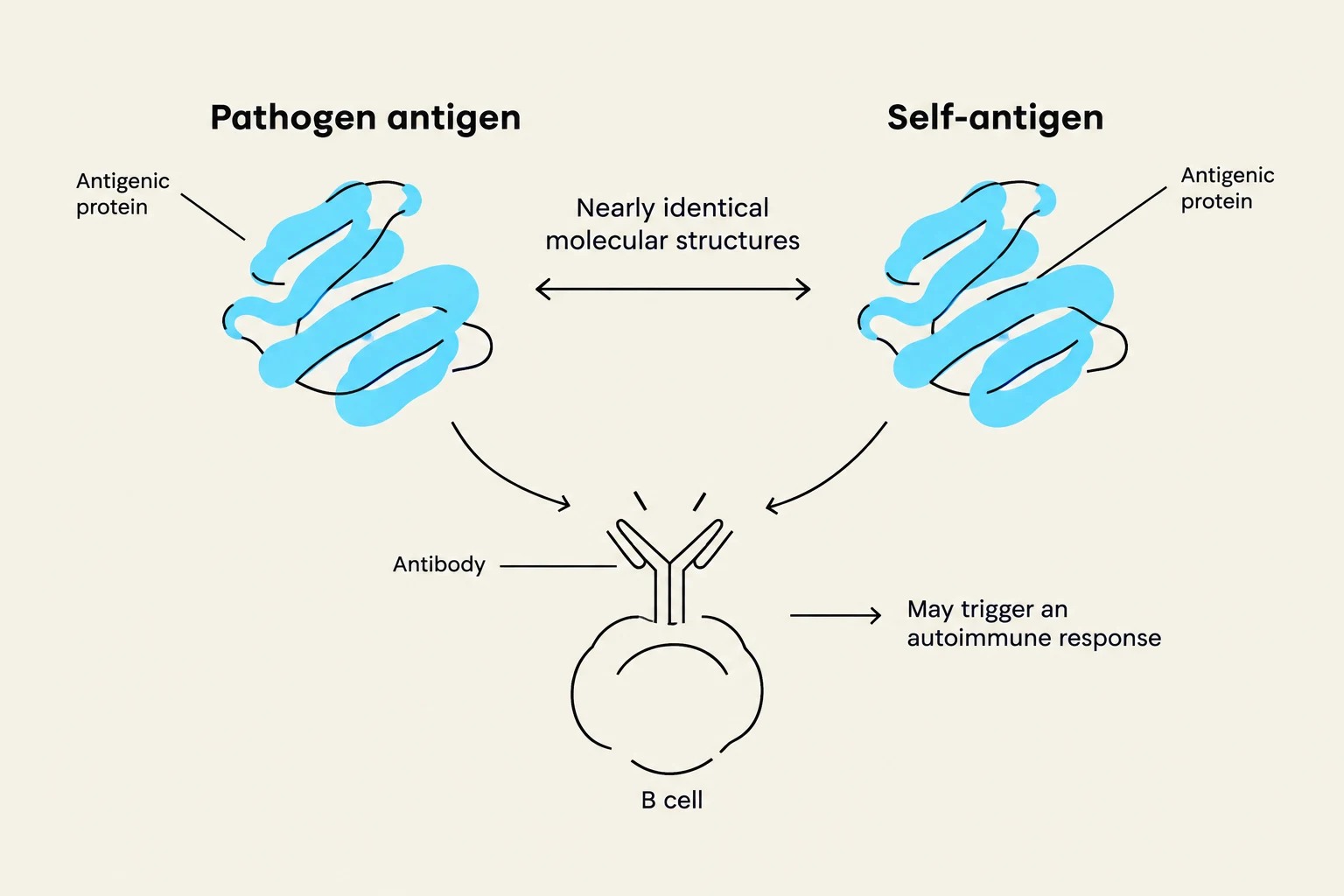
Molecular mimicry occurs when a foreign pathogen (such as a bacterium or virus) possesses structural features that look very similar to the body's own molecules. When the immune system mounts a vigorous attack against the pathogen, the resulting antibodies or T cells cannot distinguish between the germ and the host's tissue.
This "cross-reactivity" means the immune response intended for the infection inadvertently turns against the body. Rheumatic fever is a primary example of this phenomenon, where an infection triggers an immune response that subsequently attacks heart tissue due to structural similarities.
This diagram illustrates how antibodies can react to both pathogen antigens and self-antigens due to similar molecular structures, potentially leading to an autoimmune response.
3. Inappropriate Expression of Class II MHC Molecules
In a standard immune response, Class II MHC molecules are primarily found on professional antigen-presenting cells (APCs), such as macrophages or B cells. These molecules are responsible for "presenting" processed antigens to Helper T (TH) cells to initiate an immune response.
Under certain pathological conditions, somatic cells (regular body cells) may inappropriately express these Class II MHC molecules on their surfaces. This allows the body's own cells to present self-peptides directly to T cells, effectively "inviting" an autoimmune attack. This mechanism is specifically associated with the development of diabetes mellitus.
4. Nonspecific Activation of B Cells
The fourth mechanism involves a breakdown in the regulatory checkpoints that control B cell activation. Normally, B cell activation is a highly specific process involving antigen binding and subsequent cytokine signaling from Helper T cells.
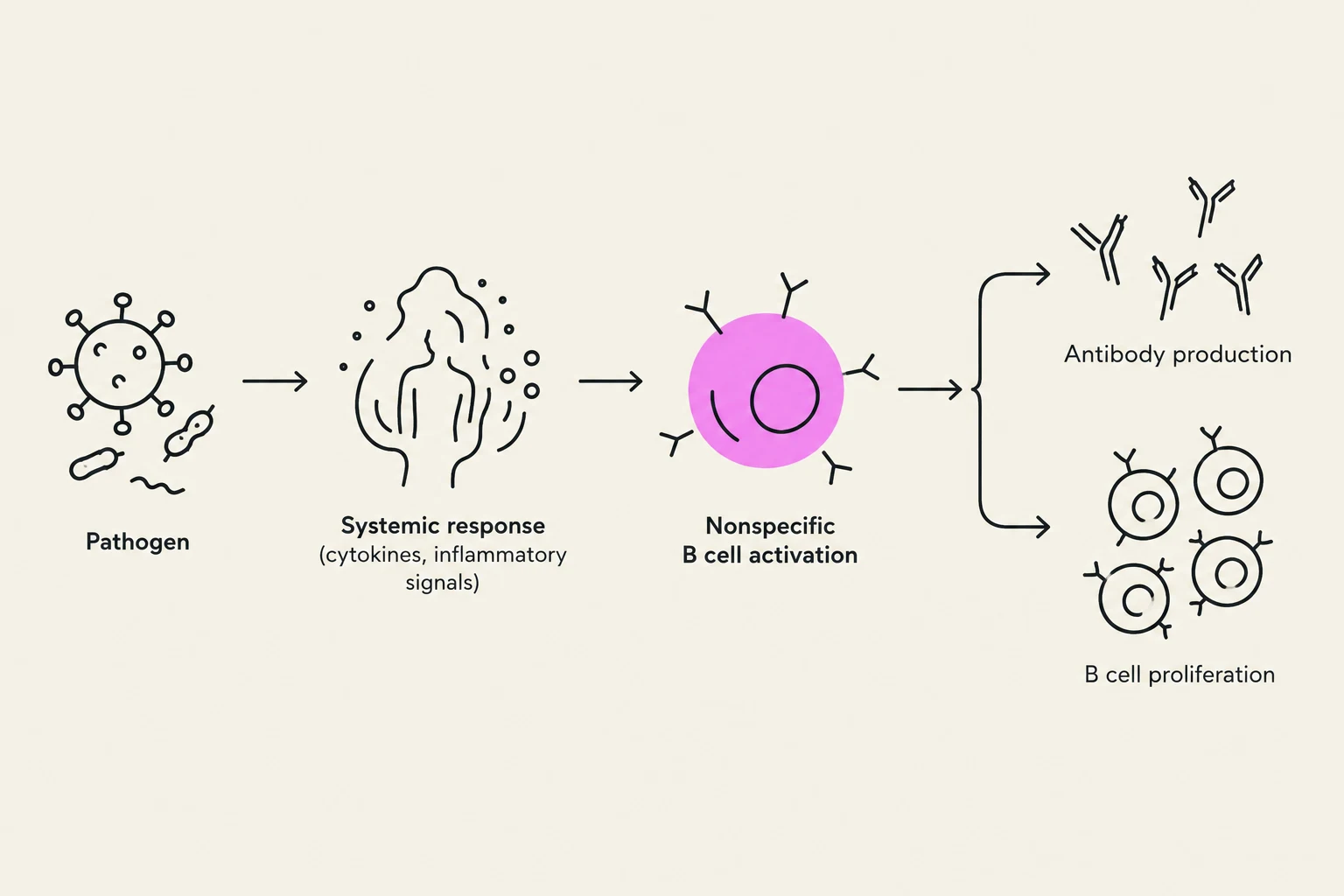
However, certain pathogens can trigger a massive, systemic immune response that causes the nonspecific activation of B cells. This bypasses the usual "checks and balances," leading to the production of various autoantibodies that circulate through the body and attack multiple systemic components. Systemic lupus erythematosus (SLE) is a hallmark disorder driven by this type of widespread, nonspecific activation.
This diagram illustrates the immune response to a pathogen, showing systemic response, nonspecific B cell activation, antibody production, and B cell proliferation.
Summary Table of Autoimmune Mechanisms
Mechanism
Description
Clinical Example
Antigen Release
Hidden self-antigens are exposed to the immune system for the first time.