Explore the physical principles of pressure and volume that drive breathing, the anatomy of the lungs including alveoli and pleurae, and the mechanics of inspiration and expiration. Learn about lung pressures, elasticity, and the role of surfactant in efficient gas exchange.
Breathing is a dynamic process driven by the physical principles of pressure and volume. To move air into and out of the body, the respiratory system must create pressure gradients between the atmosphere and the lungs. This article explores how the anatomy of the lungs, the physics of gas laws, and the specialized structures of the respiratory system work in harmony to facilitate efficient gas exchange.
Alveoli and the Pathway to Red Blood Cells
The functional unit of the respiratory system is the alveolus—a cluster of thin-walled, grape-like sacs. These sacs are the primary site of gas exchange. Two specialized cell types line the alveoli: Type I alveolar cells, which cover about 95% of the surface and form the thin wall for gas exchange, and Type II alveolar cells, which cover 5% of the surface and secrete pulmonary surfactant.
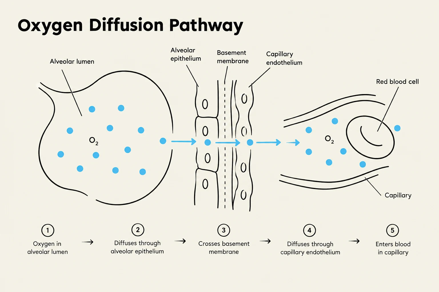
For oxygen to reach the hemoglobin inside a red blood cell (RBC), it must traverse a specific sequence of layers:
The Alveolar Lumen (connected to the outside environment)
The water/surfactant layer
The Type I alveolar cell membrane
The basement membrane of the alveoli
The interstitial fluid
The capillary basement membrane
The endothelial cell of the capillary
The blood plasma
The RBC membrane
The RBC cytoplasm (where hemoglobin resides)
Concept Check: Optimal Gas Exchange
Efficient gas exchange depends on a short diffusion distance. Factors like pulmonary edema (excess fluid) or scar tissue can increase this distance, impairing exchange. Additionally, adequate airflow (maintained by airway diameter) and sufficient blood flow are critical for maintaining optimal exchange.
Diagram illustrating the pathway of oxygen diffusion from the alveolar lumen into the bloodstream, passing through the alveolar epithelium, basement membrane, and capillary endothelium.
Pleurae and the Mechanics of Breathing
The lungs are not physically attached to the thoracic wall; instead, they are housed within the pleural sac, a closed, double-walled sac. The intrapleural space between these walls is filled with a tiny amount of intrapleural fluid. This fluid serves two vital purposes: it allows the pleurae to slide past one another during breathing, and its cohesiveness (the attraction between polar water molecules) helps hold the lungs against the thoracic wall.
The mechanics of breathing rely on Boyle’s Law, which states that pressure and volume are inversely related:
P1V1=P2V2
When the volume of the thoracic cavity increases, the pressure within the airways decreases, and vice versa.
Inspiration and Expiration
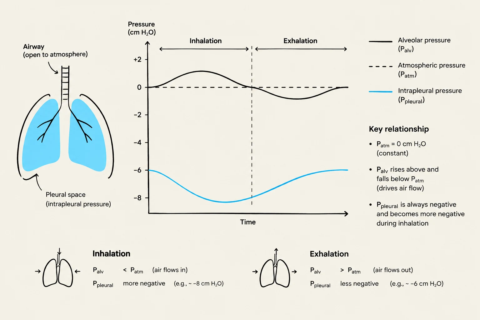
Inspiration: The diaphragm and external intercostal muscles contract, enlarging the thoracic cavity. This increase in volume causes the alveolar pressure (PA) to drop below atmospheric pressure (PB). Air flows down this pressure gradient into the lungs.
Expiration: The inspiratory muscles relax, and the elastic recoil of the lungs and chest wall decreases the thoracic volume. This decrease in volume raises the alveolar pressure (PA) above atmospheric pressure (PB), forcing air out of the lungs.
Lung Pressures and the Transmural Gradient
To understand how the lungs stay inflated, we must distinguish between different types of pressure:
PB: Barometric (atmospheric) pressure.
PA: Alveolar pressure.
PIP: Intrapleural pressure.
PTP: Transpulmonary (transmural) pressure.
The transpulmonary pressure gradient is defined as:
PTP=PA−PIP
This gradient represents the difference between the pressure inside the lung and the pressure in the pleural space. It acts as an outward force that opposes the natural inward elastic recoil of the lungs, effectively keeping them distended and open.
At rest, the intrapleural pressure (PIP) is subatmospheric (negative). This occurs because the lungs want to recoil inward while the thoracic wall wants to recoil outward, pulling the pleural linings apart and creating a vacuum-like effect.
This diagram illustrates the pressure changes in the lungs during inhalation and exhalation, showing the relationship between alveolar pressure, atmospheric pressure, and intrapleural pressure.
Pulmonary Elasticity and Surfactant
The lungs possess elasticity, which is a combination of elastic connective tissue and alveolar surface tension. This leads to two key properties:
Compliance (C): A measure of how easily the lungs can be stretched. It is calculated as:
C=ΔPΔV
Elastic Recoil: The tendency of the lungs to return to their original size, driven largely by surface tension.
Alveolar Surface Tension is caused by the strong attraction between water molecules at the air-water interface within the alveoli. Without intervention, this tension would cause the alveoli to shrink or even collapse. According to Laplace’s Law, the inward collapsing pressure (P) is proportional to surface tension (T) and inversely proportional to the radius (r) of the alveolus:
P=r2T
This means smaller alveoli have a much greater tendency to collapse than larger ones. To prevent this, Type II alveolar cells secrete pulmonary surfactant. Surfactant is composed of roughly 90% lipids and 10% proteins. By interfering with the interaction between water molecules, surfactant reduces surface tension, increases lung compliance, and stabilizes alveolar size, making the lungs much easier to inflate.
Exam Tip: The Bohr and Haldane Effects
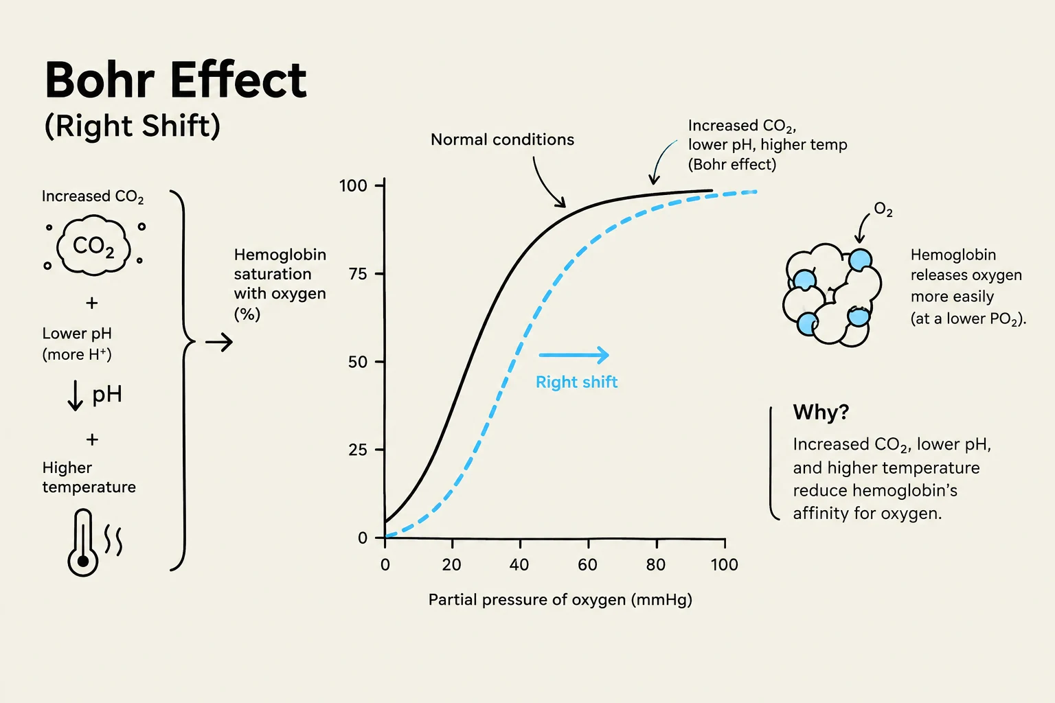
Remember that these two effects work in synchrony to optimize gas transport. The Bohr Effect describes how increased CO2 and acidity ($H^+$) decrease hemoglobin's affinity for O2, helping unload oxygen at the tissues. The Haldane Effect describes how the unloading of O2 facilitates the pickup of CO2 at the tissues.
The Bohr effect illustrates how changes in blood CO2, pH, and temperature affect hemoglobin's affinity for oxygen. An increase in CO2, a decrease in pH, and an increase in temperature shift the oxygen-hemoglobin dissociation curve to the right, indicating that hemoglobin releases oxygen more readily.
Keep reading for free.
Create an account to finish this reading and make it part of your study routine.