


Understanding the internal anatomy of the dentino-pulpar complex is the cornerstone of predictable endodontic treatment. Success depends on the clinician's ability to visualize the pulp chamber's size, shape, and the number, location, and curvature of the canals before the first cut is made. For the maxillary anterior teeth, this requires a precise understanding of root length, canal prevalence, and the specific anatomical landmarks that dictate safe access depth.
The Maxillary Anterior Teeth: Morphology and Access
In the maxillary arch, the anterior teeth present relatively predictable canal configurations, but they possess unique morphological variations that can lead to complications if ignored.
Maxillary Central Incisor
The maxillary central incisor is almost invariably single-rooted and single-canaled, with a single canal present in $99.9%$ of cases. It has an average root length of .
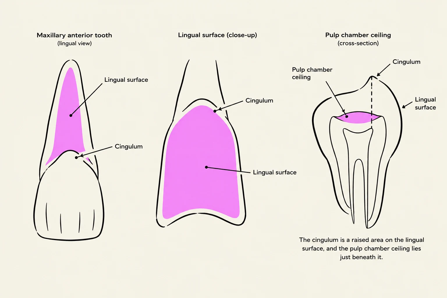
When performing access, the entry point is not on the cingulum; rather, it is located in the middle of the lingual surface, midway between the cingulum and the incisal edge. This is because the chamber ceiling sits more incisally than the cingulum. The average access depth from the surface to the chamber floor is approximately .
Maxillary Lateral Incisor
The maxillary lateral incisor is often considered the most variable anterior tooth. While it also features a single root and a single canal in $99.9%$ of cases, it is the most frequent site for several anatomical anomalies:
- Distal Dilaceration: The apex frequently shows a curve toward the distal.
- Dens Invaginatus: Also known as "dens in dente," this is an invagination of the enamel organ into the dental papilla before calcification, appearing as a "tooth within a tooth".
- Palatogingival (Palatoradicular) Groove: A developmental groove on the palatal surface.

Keep reading for free.
Create an account to finish this reading and make it part of your study routine.
Continue reading freeAlready have an account? Log in
Updated