The transition from theoretical knowledge to clinical practice in Obstetrics and Gynecology (OBGYN) requires a firm grasp of both physiological changes and the management of pregnancy-related complications. This module focuses on the continuum of care, beginning with the optimization of health before conception, through the complexities of antepartum management, and into the urgent evaluation of obstetric emergencies such as ectopic pregnancy and spontaneous abortion.
Maternal-Fetal Physiology and Preconception Care
Successful pregnancy outcomes are often determined long before conception occurs. Preconception care is a proactive approach designed to identify and modify biomedical, behavioral, and social risks to a woman's health or reproductive function that may have an impact on pregnancy.
A comprehensive preconception assessment must include a detailed history covering medical, surgical, gynecological, genetic, and psychosocial factors, as well as substance use and environmental exposures.
Key Recommendations for Optimization
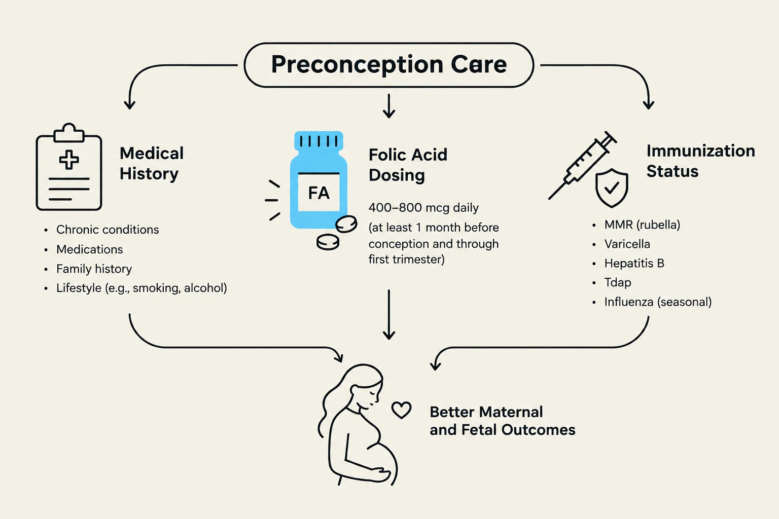
Folic Acid Supplementation: To prevent neural tube defects (NTDs), patients should take 0.4 mg of folic acid daily. However, if a patient is taking medications that interfere with folate metabolism, a higher dose of 4 mg is required.
Immunizations: Patients should be up-to-date on vaccinations for MMR (Measles, Mumps, Rubella), varicella, pertussis, and hepatitis B.
Clinical Pearl: Live vaccines, such as the MMR vaccine, must be avoided during pregnancy. Patients should ideally avoid conception for at least one month after receiving a live vaccine.
Genetic Screening: Screening should be tailored to the patient's ethnic background to identify carrier status for specific conditions:
Thalassemia: Mediterranean and Southeast Asian descent.
Tay-Sachs and Canavan disease: Ashkenazi Jews.
Cystic Fibrosis: Caucasians of European and Ashkenazi descent.
This diagram outlines key components of preconception care, including medical history, folic acid dosing, and immunization status, all contributing to better maternal and fetal outcomes.
Pregnancy Dating and Antepartum Management
Once pregnancy is confirmed, accurate dating and routine monitoring become the pillars of antepartum care.
Gestational Age and Delivery Calculation
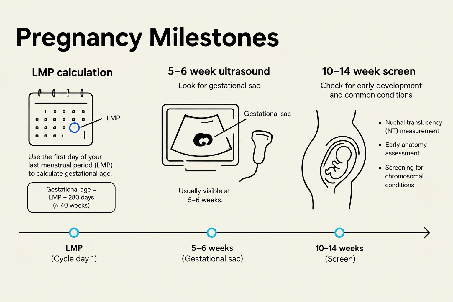
The Estimated Date of Confinement (EDC), or estimated date of delivery, is calculated using Naegele's Rule:
EDC=LMP+7 days−3 months
where LMP is the first day of the Last Menstrual Period.
Ultrasound and β-hCG Monitoring
Ultrasound (US) is a vital tool for confirming intrauterine pregnancy and assessing fetal well-being. A normal gestational sac can typically be visualized via ultrasound 5–6 weeks after the LMP.
If a patient presents with a positive pregnancy test but an inconclusive ultrasound, clinicians must consider the β-hCG level. The ability to visualize a sac is dependent on reaching specific β-hCG thresholds; if the level is low, a lack of visible sac does not necessarily rule out a viable pregnancy.
The preferred mid-pregnancy ultrasound occurs at 18–20 weeks of gestation. This scan is used to:
Confirm intrauterine pregnancy and estimate gestational age.
Evaluate for multiple gestations.
Confirm fetal cardiac activity.
Diagnose uterine abnormalities or fetal structural defects.
Routine Testing
At the first prenatal visit, routine screening for infectious diseases is mandatory to prevent vertical transmission and manage potential maternal-fetal impacts.
This illustration outlines key pregnancy milestones, including LMP calculation, 5-6 week ultrasound for gestational sac detection, and the 10-14 week screen for early development and common conditions.
Genetic Testing and Diagnostic Procedures
For patients interested in prenatal diagnosis, clinicians must distinguish between screening tests (which assess risk) and diagnostic tests (which provide definitive answers).
Test
Type
Timing
Key Information
Integrated Screen
Screening
10–22 weeks
Combines Nuchal translucency, PAPP-A, and Quad screen for higher accuracy.
Cell-Free DNA
Screening
≥10 weeks
High sensitivity for Trisomy 21, 18, 13, and sex chromosome aneuploidy.
Chorionic Villus Sampling (CVS)
Diagnostic
>10 weeks
Provides definitive diagnosis in the first trimester; carries a small risk of pregnancy loss.
Amniocentesis
Diagnostic
15–20 weeks
99% accurate for Neural Tube Defects (NTD) and structural defects.
Exam Tip: If a patient has a history of a child with a Neural Tube Defect (NTD), they require higher folic acid doses (4 mg) and should be offered amniocentesis for definitive diagnosis.
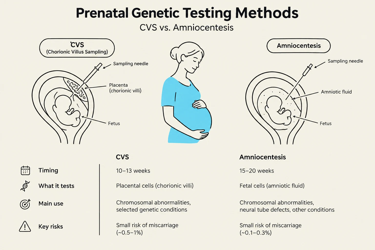
Comparison of CVS and Amniocentesis prenatal genetic testing methods, detailing their timing, what they test, main uses, and key risks.
Obstetric Emergencies: Ectopic Pregnancy and Spontaneous Abortion
First-trimester bleeding is a common clinical presentation that requires rapid differentiation between several serious conditions.
Ectopic Pregnancy
An ectopic pregnancy occurs when a fertilized egg implants outside the uterus, most commonly in the fallopian tube. Clinical suspicion is high when β-hCG levels do not correlate with ultrasound findings (e.g., a thickened endometrium without a visible intrauterine sac) or when an adnexal mass is present.
Medical Management: Methotrexate can be used for asymptomatic, motivated patients.
Contraindications to Medical Management: Known hypersensitivity to methotrexate, active pulmonary disease, peptic ulcer disease, or evidence of an immunodeficiency.
Surgical Emergency: If a patient develops severe abdominal pain, lightheadedness, tachycardia (e.g., 130 bpm), or hypotension (e.g., 90/50 mmHg), clinicians must suspect a ruptured ectopic pregnancy, which requires immediate surgical consultation.
Spontaneous Abortion
Spontaneous abortion (miscarriage) is the most common cause of early pregnancy bleeding. The clinical presentation depends on the type:
Inevitable Abortion: Characterized by vaginal bleeding and an open cervix.
Threatened Abortion: Bleeding occurs, but the cervix remains closed.
Management may include expectant management (spontaneous resolution), medical management with prostaglandins, or surgical intervention (D&C).
Clinical Note: For any patient experiencing pregnancy loss, clinicians must check the mother's blood type. If she is Rh-negative, administration of RhoGAM is essential to prevent isoimmunization.
Keep reading for free.
Create an account to finish this reading and make it part of your study routine.