Ventricular Tachycardia (VT) is a life-threatening cardiac arrhythmia characterized by a rapid sequence of wide QRS complexes originating from the ventricles. Because VT can lead to sudden cardiac arrest, the first and most critical step in management is the "Intake Rule": you must immediately check for a palpable pulse. The presence or absence of a pulse dictates two entirely different emergency pathways, as a patient with a pulse and a patient without a pulse require fundamentally different treatments.
The Critical First Step: Pulse vs. No Pulse
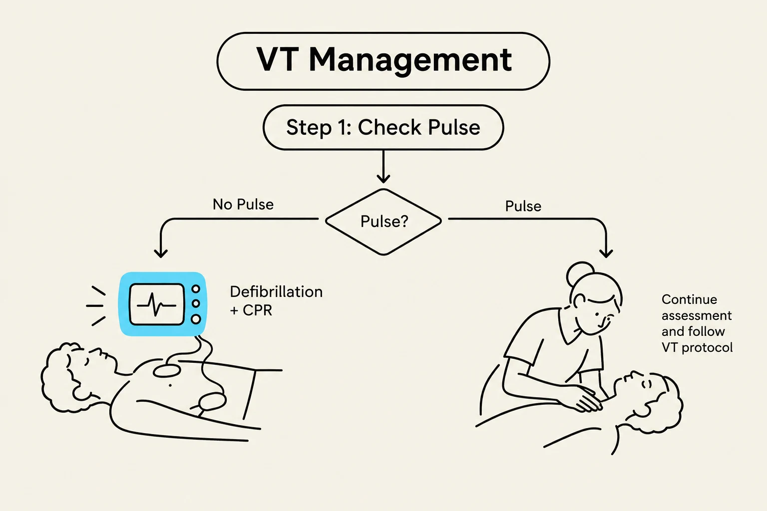
The management of VT is bifurcated by the patient's hemodynamic stability. If a patient presents with VT but has no palpable pulse, they are in a state of cardiac arrest. In this scenario, the priority is immediate CPR to act as a bridge to maintain perfusion, followed by rapid defibrillation, which is the most effective way to terminate the rhythm.
Conversely, if the patient has a pulse, the clinician must assess stability. While "stable" VT is considered rare, a patient with a pulse may present with palpitations, syncope, or chest pain.
Exam Tip: Always check the pulse first. If there is no pulse, it is a "code" situation requiring unsynchronized shock (defibrillation). If there is a pulse, you may need synchronized cardioversion.
Monomorphic Ventricular Tachycardia
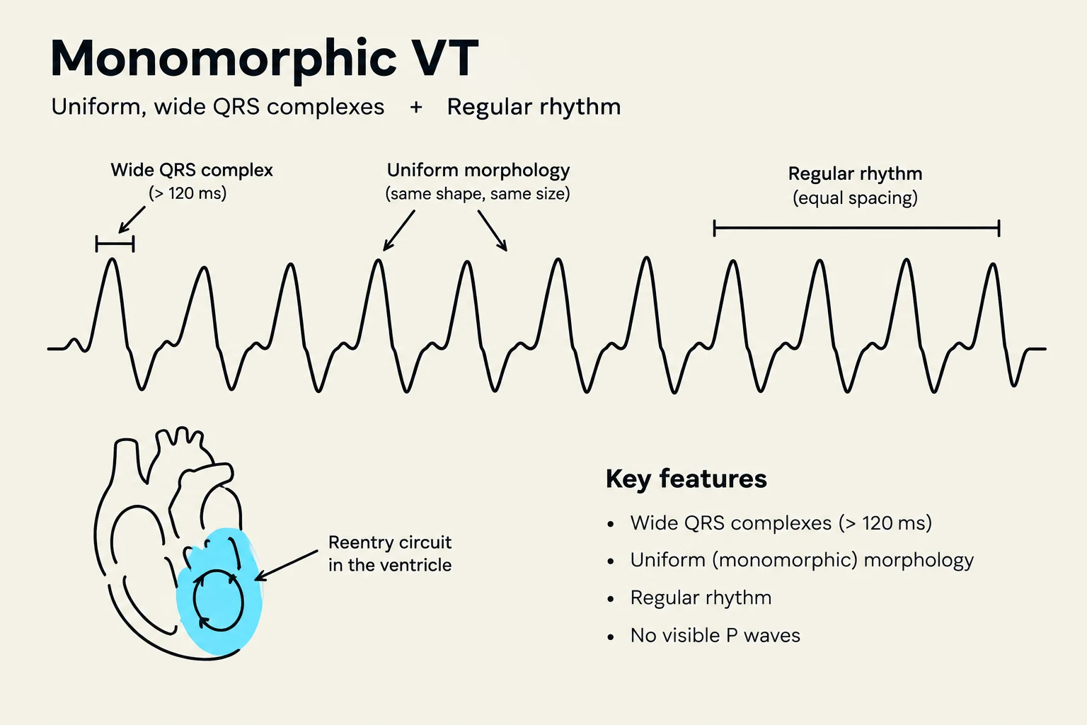
Monomorphic VT is characterized by an ECG pattern where the QRS complexes are uniform in shape and amplitude. This regularity occurs because the arrhythmia is typically driven by a single re-entrant circuit, often localized around scarred tissue from a previous myocardial infarction.
Pathophysiology and Causes
The single circuit allows the electrical impulse to repeat the same path at a high frequency. Common triggers for monomorphic VT include:
Ischemia and prior myocardial infarction (scar tissue)
Electrolyte disorders (such as hypokalemia)
Hypoglycemia and hypoxia
Acidosis or cardiac tamponade
Clinical Management
For a patient with monomorphic VT who maintains a pulse, the preferred treatment is synchronized cardioversion. Synchronization ensures the shock is delivered during the QRS complex rather than the vulnerable T-wave period. If the patient is stable, monitored antiarrhythmic infusions such as amiodarone may be used.
This diagram illustrates the key features of Monomorphic VT, a type of irregular heartbeat characterized by uniform, wide QRS complexes and a regular rhythm, often caused by a reentry circuit in the ventricle.
Polymorphic Ventricular Tachycardia
Unlike the uniform appearance of monomorphic VT, Polymorphic VT presents with QRS complexes that vary significantly in shape and amplitude. Because the electrical activity is constantly changing, you cannot "synchronize" a shock to a specific QRS complex.
Torsades de Pointes
A specific and dangerous subtype of polymorphic VT is Torsades de Pointes. This rhythm is uniquely associated with a prolonged QT interval on the ECG. In Torsades, the electrical axis appears to "twist" around the isoelectric baseline.
The pathophysiology involves a prolonged QT interval that allows the next beat to trigger off the preceding T-wave, creating a cycle of instability. This is often exacerbated by:
Electrolyte imbalances: Specifically hypokalemia (low potassium) and low magnesium.
Pharmacology: Certain QT-prolonging drugs.
Congenital factors: Long QT syndrome.
Clinical Management
Treatment for polymorphic VT depends on the presence of a pulse, but the rhythm itself dictates the method of electrical therapy:
Unsynchronized Shock (Defibrillation): Because the QRS complexes are constantly changing, synchronized cardioversion is impossible; therefore, unsynchronized defibrillation must be used.
Magnesium Treatment: For Torsades de Pointes specifically, intravenous magnesium is the definitive treatment to stabilize the cardiac membrane.
Correcting Underlying Causes: It is vital to address electrolyte disorders (magnesium and potassium) and remove any offending QT-prolonging medications.
Summary Comparison Table
To aid in rapid clinical decision-making, the following table summarizes the key differences between the primary types of VT:
Feature
Monomorphic VT (with pulse)
VT without Pulse
Polymorphic VT / Torsades
ECG Appearance
Wide QRS, uniform complexes
Wide QRS, uniform complexes
Wide QRS, varying amplitude/shape
Pulse Status
Present
Absent
May or may not be present
Primary Risk
Syncope, chest pain
Sudden death
Fainting, collapse, progression to arrest
Shock Type
Synchronized cardioversion
Unsynchronized defibrillation
Unsynchronized defibrillation
Key Medication
Amiodarone
Epinephrine (during CPR)
Magnesium (for Torsades)
Key Driver
Scar tissue / Ischemia
Acute cardiac arrest
Prolonged QT / Electrolytes
Flowchart illustrating VT (Ventricular Tachycardia) management, starting with checking for a pulse. If no pulse is detected, defibrillation and CPR are indicated. If a pulse is present, assessment continues following the VT protocol.
Final Exam Summary:
Pulse determines the shock type: Pulse = Synchronized; No Pulse = Unsynchronized.
Shape determines the subtype: Monomorphic = same shape; Polymorphic = changing shape.
Torsades = Polymorphic + Prolonged QT.
Magnesium is the drug for Torsades.
Keep reading for free.
Create an account to finish this reading and make it part of your study routine.