This content provides a comprehensive overview of gastritis and peptic ulcer disease (PUD), covering their definitions, causes, clinical manifestations, risk factors, and management strategies. It details the differences between acute and chronic gastritis, the role of H. pylori infection and NSAIDs in PUD, and essenti
Gastric and duodenal disorders represent a significant spectrum of gastrointestinal pathology, ranging from acute mucosal irritation to chronic, life-threatening ulcerations and malignancies. At the core of these conditions is the disruption of the mucosal barrier, which normally serves as a protective shield for the stomach tissue against highly acidic digestive juices. Understanding the distinction between acute and chronic presentations, as well as the role of infectious and lifestyle factors, is essential for effective clinical assessment and nursing management.
Understanding Gastritis
Gastritis is defined by the inflammation of the gastric mucosa. It is categorized based on the onset and duration of the inflammatory process.
Acute Gastritis
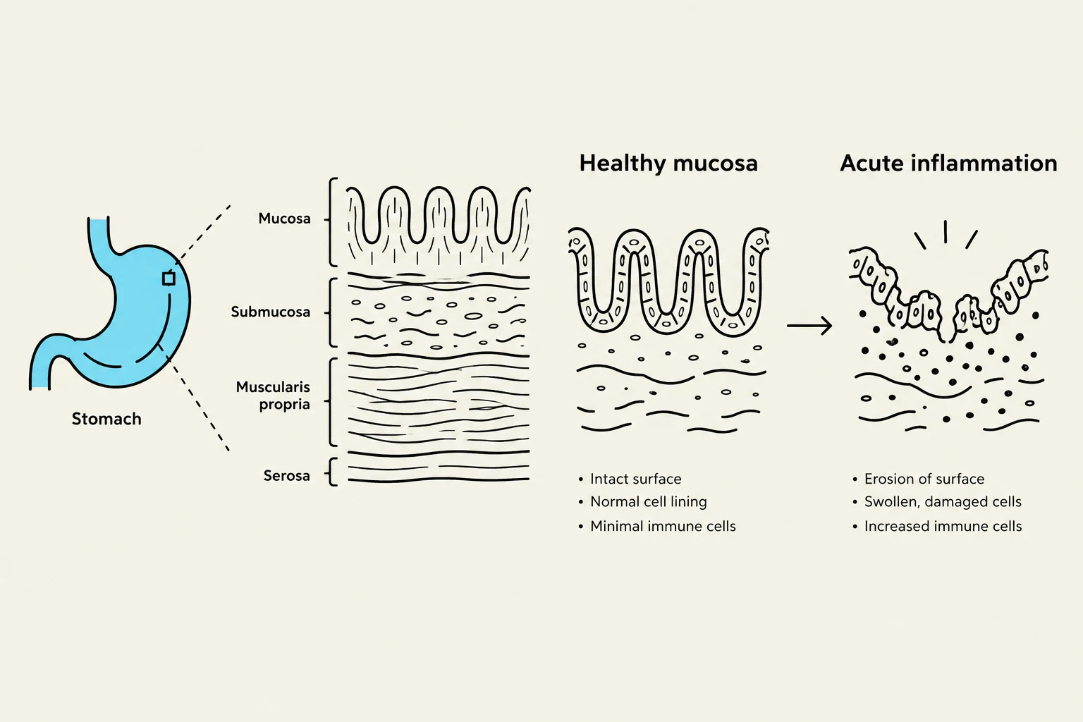
Acute gastritis is characterized by a rapid onset of symptoms. It is frequently caused by dietary indiscretion, but other significant triggers include the use of certain medications (such as NSAIDs), alcohol consumption, bile reflux, and radiation therapy. In severe cases, the ingestion of strong acids or alkalis can lead to serious complications.
Chronic and Erosive Gastritis
Chronic gastritis involves prolonged inflammation and can lead to the atrophy of gastric tissue. This condition may be caused by benign or malignant ulcers, Helicobacter pylori infection, or autoimmune diseases. Lifestyle factors such as smoking, alcohol, and chronic reflux of bile or pancreatic secretions also contribute to its progression.
Erosive gastritis is a specific, more severe form where the mucosal lining is actively worn away.
Diagram illustrating the layers of the stomach lining in a healthy state versus acute inflammation, showing cellular changes and surface erosion.
Clinical Manifestations
The symptoms of gastritis vary significantly depending on whether the condition is acute or chronic:
Acute Manifestations: Patients often present with epigastric pain, dyspepsia, anorexia, hiccups, nausea, and vomiting. If the gastritis is erosive, clinicians must monitor for signs of bleeding, such as melena (dark, tarry stools), hematemesis (vomiting blood), or hematochezia (bright red blood per rectum).
Chronic Manifestations: Symptoms may include fatigue, pyrosis (heartburn), belching, a sour taste in the mouth, halitosis, early satiety, nausea, and vomiting. Notably, chronic gastritis can lead to pernicious anemia due to the malabsorption of Vitamin B12. Some patients may remain entirely asymptomatic.
Exam Tip: When assessing a patient with suspected chronic gastritis, always check for signs of Vitamin B12 deficiency (such as neurological changes or macrocytic anemia), as this is a common complication of malabsorption.
Peptic Ulcer Disease (PUD)
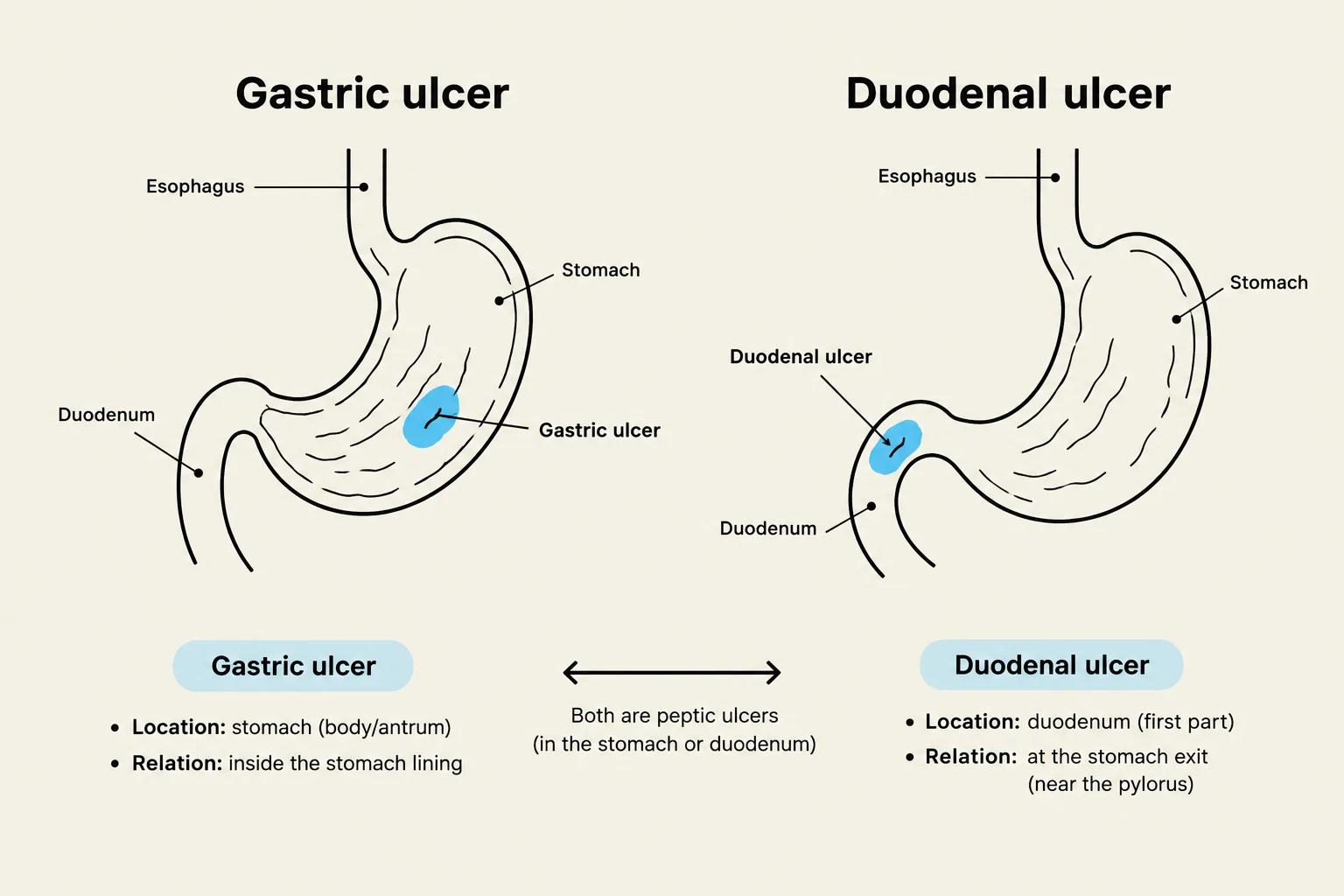
Peptic ulcer disease occurs when an erosion of the mucous membrane forms an excavation (an ulcer) in the stomach, pylorus, duodenum, or esophagus.
Risk Factors and Pathophysiology
A primary driver of PUD is infection with Helicobacter pylori. However, several other risk factors increase the likelihood of developing ulcers, including:
Excessive secretion of stomach acid
Chronic use of Non-Steroidal Anti-Inflammatory Drugs (NSAIDs)
Smoking and alcohol consumption
Dietary factors and familial tendency
Patients typically report a dull, gnawing, or burning pain in the midepigastrium, often accompanied by heartburn and vomiting.
Diagrams illustrating the locations of gastric and duodenal ulcers within the human digestive system, highlighting their respective positions in the stomach and duodenum.
Clinical Assessment and Management
Patient Assessment
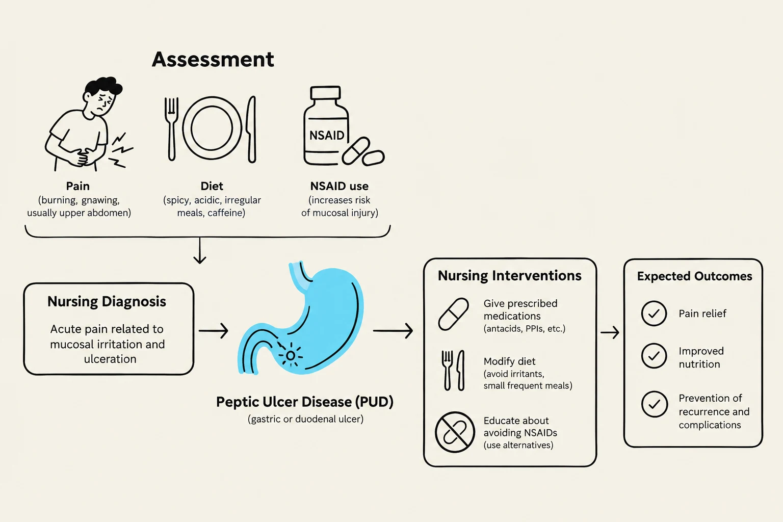
Effective management begins with a thorough assessment of the patient's history and physical status:
History: Identify presenting signs, symptoms, and a dietary history. A 72-hour food diary can help identify predictable patterns of pain related to food intake.
Medication Review: Specifically assess for the use of NSAIDs.
Physical Assessment: Conduct abdominal assessments and monitor vital signs.
Bleeding Screen: Look for signs of anemia or active bleeding.
Medical Management
The goals of medical therapy are to reduce acid production and allow the mucosa to heal:
For Acute Gastritis: Patients may need to refrain from food and alcohol until symptoms subside. Supportive therapy includes IV fluids, nasogastric intubation, antacids, histamine-2 receptor antagonists, and proton pump inhibitors (PPIs).
For Chronic Gastritis and PUD: Management focuses on lifestyle modifications (diet, stress reduction, avoiding alcohol/NSAIDs) and pharmacologic therapy. Surgery may be required in certain cases.
Nursing Management and Interventions
Nursing care focuses on pain relief, nutritional support, and the prevention of life-threatening complications.
Core Nursing Interventions
Pain and Anxiety Management: Use a calm approach to explain all procedures and treatments to reduce patient anxiety. Implement prescribed medications and dietary changes to relieve pain.
Nutritional Support: For acute gastritis, the patient should initially remain NPO (nothing by mouth). As tolerated, introduce clear liquids and then solid foods as prescribed. It is vital to discourage caffeinated beverages, alcohol, and smoking.
Fluid and Electrolyte Balance: Monitor Intake and Output (I&O) closely for signs of dehydration, electrolyte imbalances, or hemorrhage.
Patient Education: Provide counseling for smoking cessation and alcohol reduction.
Monitoring for Complications
Nurses must be vigilant for the following serious complications of PUD and gastritis:
Hemorrhage: Indicated by hematemesis, melena, or a drop in blood pressure and increase in heart rate.
Perforation and Penetration: A sudden, severe abdominal pain that may indicate the ulcer has breached the stomach wall.
Gastric Outlet Obstruction: Often caused by scarring or edema from chronic inflammation, preventing food from passing into the duodenum.
This diagram illustrates the assessment, nursing diagnosis, interventions, and expected outcomes for Peptic Ulcer Disease (PUD).
Summary Table: Management Goals
Goal
Nursing Action
Relief of Pain
Administer antacids/PPIs; modify diet
Nutritional Status
NPO for acute; gradual reintroduction of solids
Anxiety Reduction
Calm approach; clear explanation of procedures
Complication Prevention
Monitor I&O, vital signs, and stool for blood
Keep reading for free.
Create an account to finish this reading and make it part of your study routine.