Management of Cyanotic Congenital Heart Defects: Tetralogy of Fallot and Pulmonary Atresia
This guide details the management of cyanotic congenital heart defects, including Tetralogy of Fallot (TOF), pulmonary atresia, and absent pulmonary valve syndrome. It covers clinical presentations, hypercyanotic spells, surgical interventions like palliative shunts and complete repair, and long-term follow-up, includi
Cyanotic congenital heart defects present a significant challenge in pediatric cardiology, requiring a nuanced understanding of anatomy, hemodynamics, and surgical timing. The management of these conditions—ranging from Tetralogy of Fallot (TOF) to more complex presentations like pulmonary atresia—focuses on relieving pressure gradients, maintaining systemic perfusion, and ensuring long-term ventricular function through medical, interventional, or surgical means.
Tetralogy of Fallot (TOF)
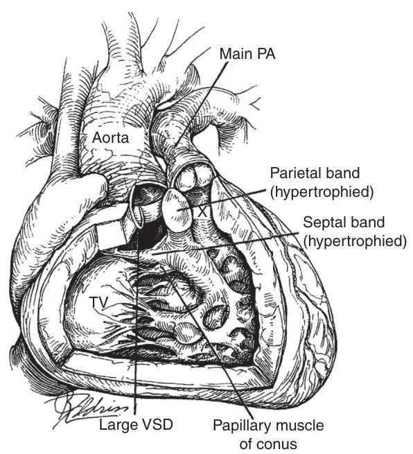
Tetralogy of Fallot is a classic cyanotic defect characterized by four hallmark anatomical findings: an outlet-type ventricular septal defect (VSD), right ventricular outflow tract (RVOT) obstruction, an overriding aorta, and right ventricular hypertrophy (RVH).
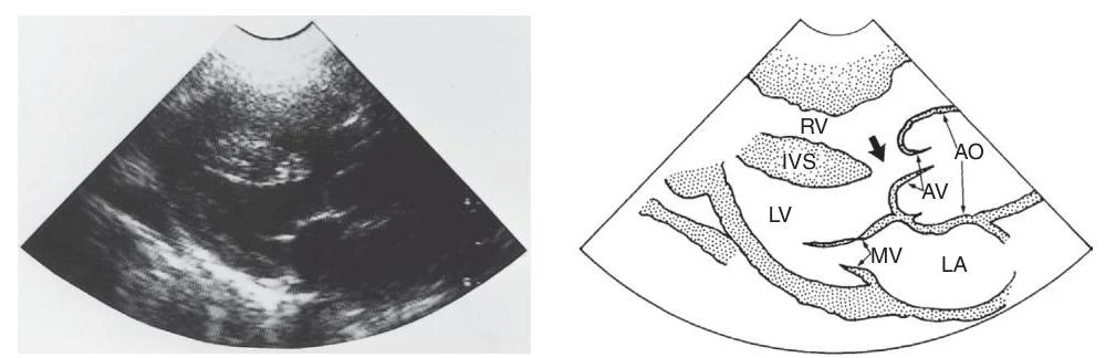
An illustration of a heart showing a large ventricular septal defect (VSD) with hypertrophied parietal and septal bands, and a prominent papillary muscle of the conus. The aorta and main pulmonary artery (Main PA) are also labeled.Diagram of the heart chambers and valves, including the right ventricle (RV), interventricular septum (IVS), left ventricle (LV), left atrium (LA), aortic valve (AV), and mitral valve (MV).
Clinical Presentation and Hypercyanotic Spells
Infants with TOF may present with varying degrees of cyanosis. A critical clinical emergency in these patients is the "hypercyanotic spell" (or "Tet spell"). During these episodes, systemic vascular resistance (SVR) may drop or RVOT obstruction may increase, leading to increased right-to-left shunting across the VSD.
Management of Hypoxic Spells:
Physical Maneuvers: Squatting or "knees-to-chest" positions increase SVR, which helps reduce the right-to-left shunt.
Medical Intervention: Oxygen, morphine, and increasing SVR are primary steps. Ketamine (1–3 mg/kg) can be used to increase SVR and provide sedation, while Propranolol (0.01–0.25 mg/kg) may help reverse the spell by reducing heart rate.
Surgical Management
The goal of definitive repair is the patch closure of the VSD and the relief of RVOT obstruction.
Palliative Shunts: In specific cases—such as neonates with pulmonary atresia, hypoplastic pulmonary arteries, or unfavorable coronary anatomy—a shunt may be placed to increase pulmonary blood flow (PBF). The Modified Blalock-Taussig (BT) shunt, using a Gore-Tex interposition between the subclavian artery and the ipsilateral pulmonary artery, is the current standard.
Complete Repair: Most centers prefer elective primary repair between 3 and 12 months of age. The procedure involves VSD patch closure and widening the RVOT via infundibular resection or transannular patches.
Exam Tip: When discussing TOF repair, remember that surgeons aim to avoid a transannular patch whenever possible to prevent long-term pulmonary regurgitation. If the pulmonary annulus is too small, a transannular patch becomes unavoidable.
Long-term Follow-up
Lifelong follow-up is mandatory. A major long-term complication is pulmonary regurgitation (PR). If PR becomes moderate to severe (regurgitation fraction ≥25%), it can lead to RV volume overload and dysfunction. In such cases, pulmonary valve replacement (PVR) via transcatheter (e.g., Melody or SAPIEN valves) or surgical means may be required.
Pulmonary Atresia
Pulmonary atresia represents the extreme form of RVOT obstruction, occurring in 15–20% of TOF cases. In this condition, there is no direct connection between the RV and the pulmonary artery (PA).
Pathophysiology and Anatomy
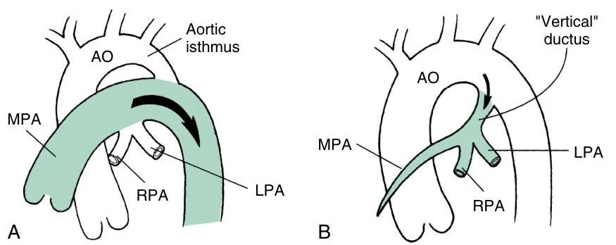
PBF in these patients is typically mediated by a patent ductus arteriosus (PDA) or through Multiple Aortopulmonary Collateral Arteries (MAPCAs).
PDA-dependent: The central PAs are usually confluent.
MAPCAs-dependent: The central PA is often nonconfluent, and the patient is designated as "pulmonary atresia and VSD" (PA-VSD).
Diagrams illustrating the aortic arch and pulmonary artery branching. Image A shows a normal configuration, while Image B depicts a variation with a "vertical" ductus.
To quantify the degree of PA hypoplasia, clinicians use the McGoon ratio and the Nakata index:
McGoon Ratio: The ratio of the sum of the diameters of the RPA and LPA to the diameter of the descending aorta.
Nakata Index: The cross-sectional area of the RPA and LPA divided by the body surface area (BSA).
Surgical Management: Unifocalization
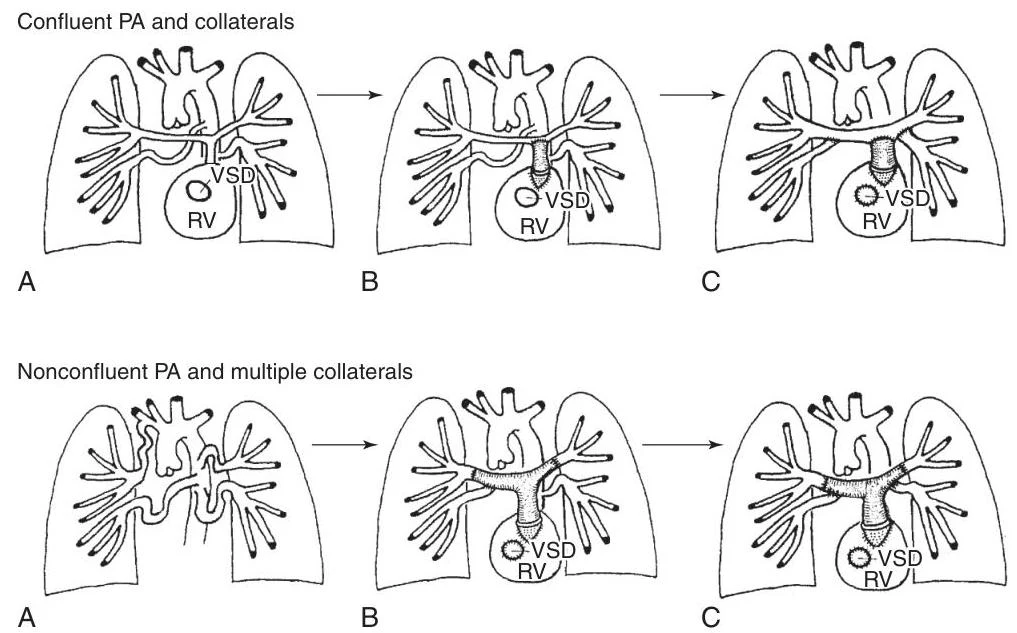
For patients with MAPCAs, a staged approach is often necessary:
Stage 1: Establishing RV-to-PA connection (e.g., via a homograft conduit) to allow central PAs to grow.
Stage 2: Unifocalization: A complex procedure where MAPCAs are divided from their aortic origins and anastomosed to the true PAs to create a single, unified pulmonary circulation.
Stage 3: Closure of the VSD.
Diagrams illustrating two types of pulmonary artery (PA) and collateral circulation in the context of a ventricular septal defect (VSD) and right ventricle (RV).
Absent Pulmonary Valve Syndrome
This rare variant ($2%$ of TOF cases) is characterized by the complete absence or rudimentary nature of the pulmonary valve leaflets.
Clinical Presentation
The hallmark is massive, aneurysmal dilatation of the pulmonary arteries due to severe pulmonary regurgitation. This dilatation can compress the lower trachea and bronchi, leading to significant respiratory distress and airway obstruction.
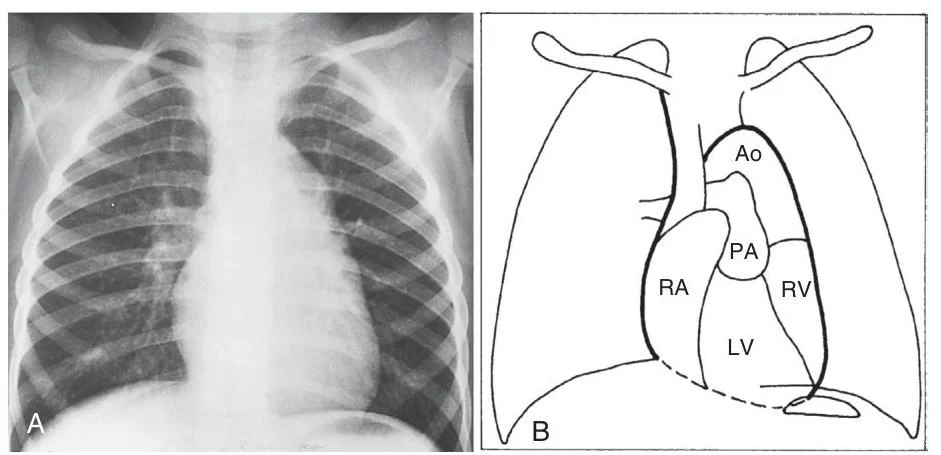
Diagram of the human heart and major blood vessels, with a corresponding chest X-ray.(Note: While the provided figure description relates to TGA, the text notes that in Absent Pulmonary Valve, the lung fields often show hyperinflated areas due to airway compression.)
Management
Surgical intervention is required for symptomatic neonates. Primary repair involves VSD closure and the use of a pulmonary homograft or valved conduit to replace the dysplastic valve and reconstruct the aneurysmal PAs.
Summary Table of Management Strategies
Condition
Primary Goal
Key Surgical/Interventional Approach
TOF
Relieve RVOT obstruction & close VSD
Primary repair (VSD patch + RVOT widening)
Pulmonary Atresia
Establish PBF
Shunt (BT) → Unifocalization → VSD closure
Absent Pulmonary Valve
Relieve airway compression
VSD closure + PA reconstruction/homograft
Keep reading for free.
Create an account to finish this reading and make it part of your study routine.