An overview of the four primary tissue types in the human body: epithelial, connective, muscle, and nervous tissues. Learn about their characteristics, classifications, and functions.
In the study of histology, a tissue is defined as a group of similar cells that work together to perform a specific function. While the human body contains trillions of cells, they are not randomly scattered; instead, they are organized into four primary tissue types: epithelial, connective, muscle, and nervous tissues. These tissues originate from three embryonic germ layers—the ectoderm, mesoderm, and endoderm—which dictate the eventual specialized roles these tissues will play in the adult body.
Epithelial Tissue: The Body's Boundary
Epithelial tissue serves as a versatile covering and lining for body surfaces, cavities, and organs. It also forms the basis of many glands. One of its most defining features is high cellularity, meaning the cells are packed very tightly together with very little extracellular material between them.
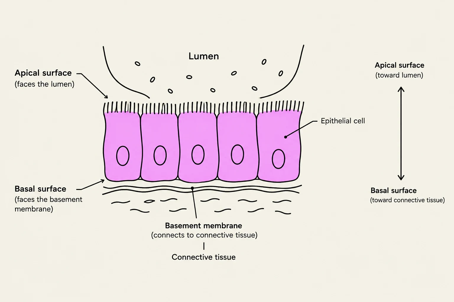
Epithelia are characterized by polarity, meaning they have two distinct sides:
Apical Surface: The upper free surface exposed to the body exterior or the cavity of an internal organ.
Basal Surface: The lower surface that is attached to the underlying connective tissue.
To maintain structural integrity and communication, epithelial cells utilize various cell junctions to adhere to one another and to each other.
Diagram illustrating the structure of epithelial cells, showing their apical and basal surfaces, basement membrane, and surrounding connective tissue.
Classification of Epithelia
To identify epithelial tissue under a microscope, scientists look at two main criteria: the number of cell layers and the shape of the cells.
1. Number of Layers:
Simple: A single layer of cells, ideal for absorption and filtration.
Stratified: Multiple layers of cells, providing protection against abrasion.
2. Cell Shapes:
Squamous: Flat and scale-like.
Cuboidal: Box-like, approximately as tall as they are wide.
Columnar: Tall and rectangular.
Specialized arrangements include pseudostratified columnar epithelium, where cells appear layered because nuclei are at different levels, but every cell actually touches the basement membrane. Another unique type is transitional epithelium, which can change shape (stretch) to accommodate volume changes, such as in the urinary bladder.
Glandular Epithelium
Epithelial tissue also forms glands. These are categorized into two functional types:
Exocrine Glands: Secrete products (like sweat or enzymes) into ducts that lead to a body surface or into a cavity.
Endocrine Glands: Ductless glands that secrete hormones directly into the bloodstream.
Exam Tip: When identifying epithelium, always look for the "free edge" (apical surface). If you can't find a free surface, you might be looking at connective tissue instead!
Connective Tissue: Support and Integration
Unlike epithelial tissue, connective tissue (CT) is characterized by having much less cellularity and more extracellular matrix. While epithelia are avascular (lacking blood vessels), most connective tissues are highly vascular, though some, like cartilage, are avascular.
The Extracellular Matrix (ECM)
The matrix is the non-living material that surrounds the cells. It consists of two main components:
Ground Substance: An amorphous, gel-like, or fluid substance that fills the space between cells and fibers.
Fibers: Protein strands that provide strength and flexibility. There are three types:
Collagen fibers: Provide high tensile strength.
Elastic fibers: Allow for stretch and recoil.
Reticular fibers: Fine, branching fibers that form a supportive "net".
Diagram illustrating the components of connective tissue matrix, including fibroblasts, ground substance, mast cells, macrophages, collagen fibers, elastic fibers, and adipocytes.
Cell Types and Variations
Connective tissue contains various cell types, often identified by suffixes. For example, immature cells often end in "-blast" (like fibroblasts), while mature cells often end in "-cyte" (like osteocytes).
Connective tissue is incredibly diverse. It ranges from the fluid blood to the rigid bone, and from the flexible hyaline cartilage to the cushioning adipose tissue (fat).
Muscle Tissue: Movement and Force
Muscle tissue is specialized for contraction, which produces movement. It is highly vascular and contains cells that are often long and cylindrical.
There are three distinct types of muscle tissue, which can be differentiated by their appearance and how they are controlled:
Muscle Type
Striated?
Voluntary?
Location
Skeletal
Yes
Yes
Attached to bones
Cardiac
Yes
No
Heart walls
Smooth
No
No
Walls of hollow organs
Skeletal muscle is "striated" (striped) and under voluntary control. Cardiac muscle is also striated but is involuntary; it features unique structures called intercalated discs and gap junctions that allow the heart to contract as a single unit. Smooth muscle is "nonstriated" and involuntary, found in the walls of organs like the stomach or blood vessels.
Nervous Tissue: Communication and Control
The final major tissue type is nervous tissue, which is responsible for receiving stimuli and transmitting electrical impulses throughout the body.
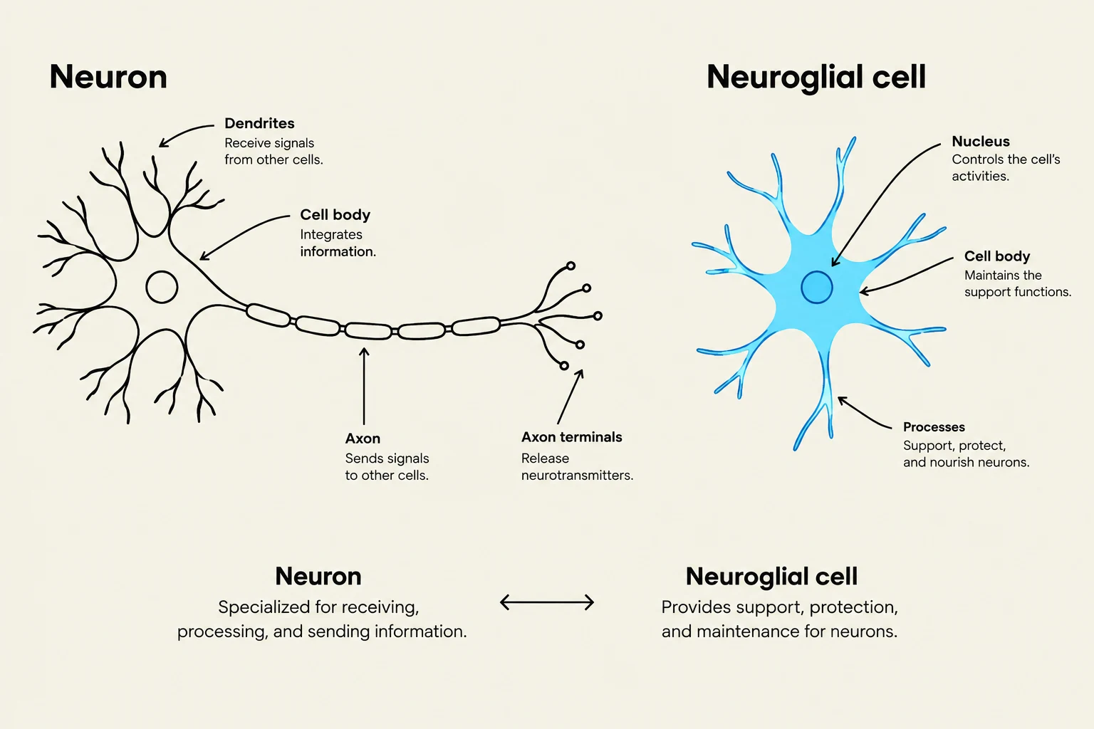
Nervous tissue is composed of two major cell types that function very differently:
Neurons: The functional units that generate and conduct electrical impulses.
Neuroglia (Glial cells): The supporting cells that protect, nourish, and insulate the neurons.
Diagram illustrating the structure and function of a neuron and a neuroglial cell.
By integrating these four tissues—the protective epithelia, the supportive connective tissues, the contractile muscles, and the communicative nervous tissue—the human body is able to maintain homeostasis and interact with its environment.
Keep reading for free.
Create an account to finish this reading and make it part of your study routine.