An overview of the vascular anatomy of the lower extremity, detailing the major arteries and veins, their flow, and common abnormalities like varicose veins and Peripheral Artery Disease (PAD).
Understanding the vascular anatomy of the lower extremity is essential for anyone studying human physiology, particularly those in the field of exercise science. The circulatory system is divided into systemic and pulmonary circuits, with the systemic circuit responsible for delivering oxygenated blood to the body's tissues and returning deoxygenated blood to the heart. While the upper extremity and head have their own complex networks, the lower extremity relies on a specific sequence of branching arteries and merging veins to support the high metabolic demands of locomotion and weight-bearing.
Major Arteries of the Lower Extremity
Arteries are classified as "resistance vessels" because they must withstand and manage high blood pressure. In the lower body, these vessels transition from large conducting arteries to smaller distributing and resistance arteries.
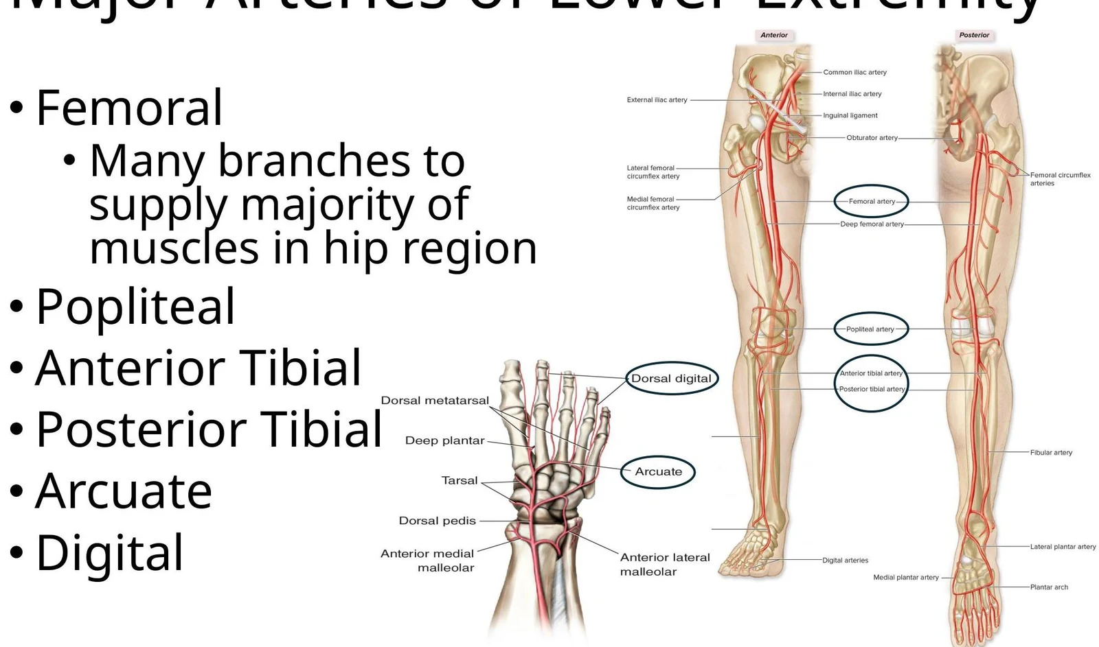
The arterial supply to the lower limb begins with the branching of the descending aorta into the common iliac arteries. From the common iliac, the vessels split into the internal and external iliac arteries. The external iliac artery is a primary conduit that eventually becomes the femoral artery, which provides extensive branching to supply the majority of the muscles in the hip region.
As the blood moves further down the limb, the arterial pathway follows a specific sequence:
Femoral Artery: Supplies the hip and upper thigh.
Popliteal Artery: Located behind the knee.
Anterior and Posterior Tibial Arteries: Supply the lower leg.
Arcuate and Digital Arteries: Provide the final distribution to the foot and toes.
Diagram illustrating the major arteries of the lower extremity, including the femoral, popliteal, anterior tibial, posterior tibial, arcuate, and digital arteries.
Arterial Flow Diagram
To visualize the progression of blood flow, one can follow the branching sequence from the core to the periphery:
Major Veins of the Lower Extremity
Unlike arteries, veins are known as "capacitance vessels" because they hold the majority of the body's blood volume. In the lower extremity, the venous system is organized into two distinct layers: deep veins and superficial veins.
Deep Veins
Deep veins are located beneath the muscle layers and typically follow the same path as their corresponding arteries. This system includes:
External iliac
Femoral and Femoral profundus
Popliteal
Anterior and Posterior tibial
Peroneal
Dorsal venous arch and Digital veins
Superficial Veins
Superficial veins reside closer to the skin surface and are distinct from the deep system. The two primary superficial veins of the lower limb are:
Greater saphenous vein: This is the longest vein in the body and is frequently utilized in medical procedures such as bypass grafting.
Lesser saphenous vein.
Diagram of the superficial veins of the leg, including the great saphenous vein, femoral vein, and dorsal venous arch.
Venous Flow Diagram
The return of blood from the lower extremity to the heart follows a merging pattern, moving from the small vessels of the foot toward the large venae cavae:
Clinical Note: Venous Return Mechanisms
Because blood in the lower extremities must fight gravity to return to the heart, the body utilizes two "pumps":
Skeletal Muscle Pump: Contraction of deep muscles compresses veins, pushing blood upward and inhibiting backflow.
Respiratory Pump: Changes in thoracic and abdominal pressure during breathing help pull blood toward the heart.
Vessel Abnormalities
Vascular health can be compromised by several conditions that affect either the arterial or venous systems.
Incompetent Valves and Varicose Veins
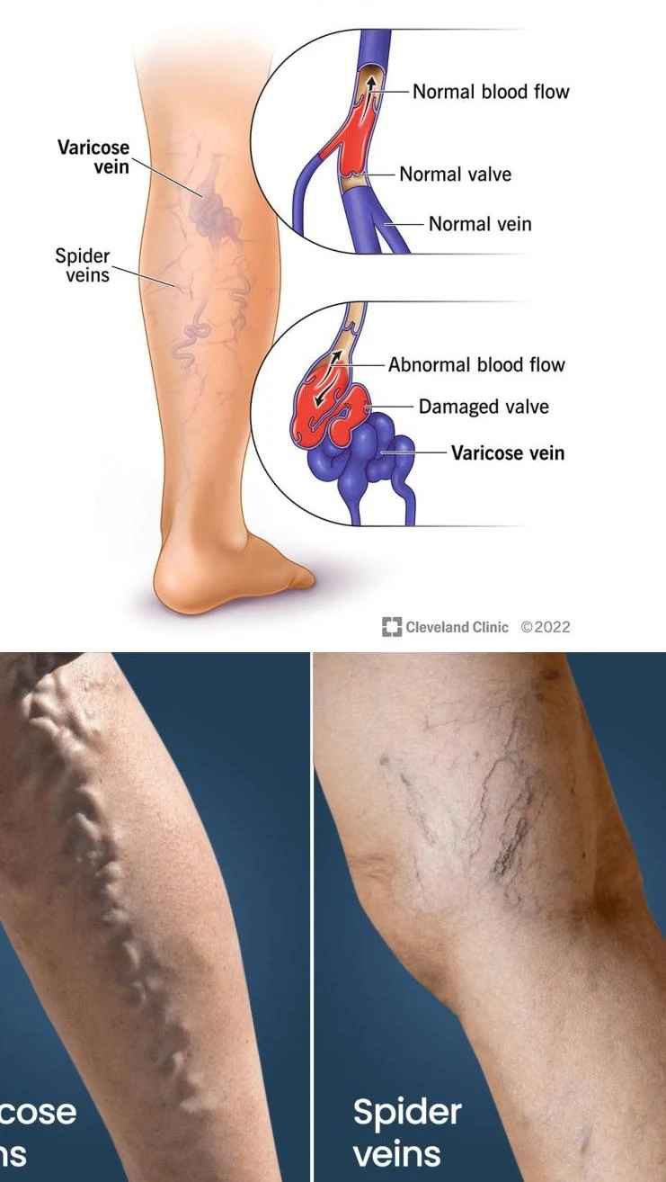
Veins contain valves to prevent the backflow of blood. When these valves become incompetent (fail to close properly), blood can pool in the vessels due to increased volume. This pooling leads to varicose veins, which are characterized by swollen, distended vessels.
Illustration comparing normal veins with normal blood flow and valves to varicose veins with abnormal blood flow and damaged valves. The image also shows real-life examples of varicose veins and spider veins on legs.
Peripheral Artery Disease (PAD)
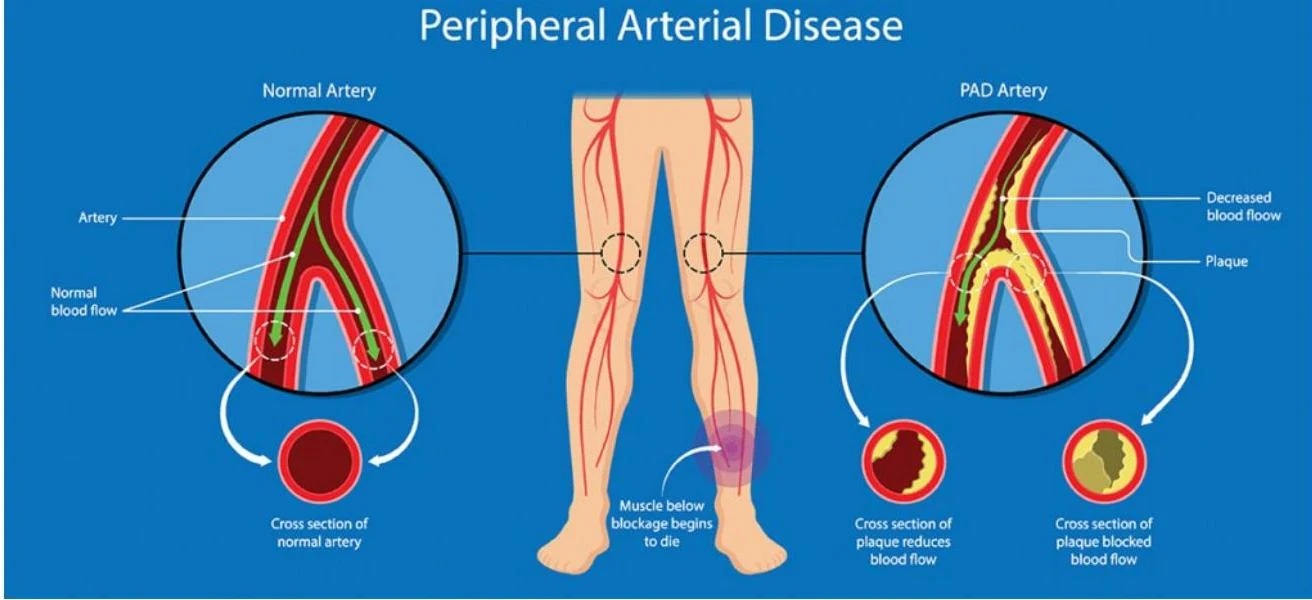
While varicose veins are a venous issue, Peripheral Artery Disease affects the arterial system. It involves the narrowing of arteries in the limbs, usually due to atherosclerosis (plaque buildup). This reduction in blood flow can cause significant pain, numbness, or cramping during physical activity.
This illustration compares a normal artery with an artery affected by Peripheral Arterial Disease (PAD), showing plaque buildup and reduced blood flow.
Important Arteries for Exercise Physiologists
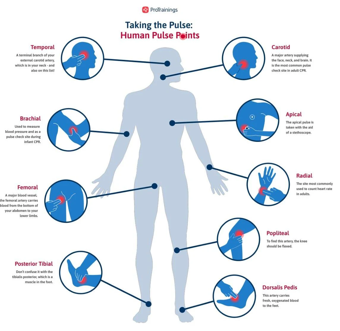
For professionals working in exercise physiology, knowing specific arterial locations is vital for monitoring client health and performance. While the lower extremity is the focus of this study, several arteries in the upper body and neck are standard clinical landmarks:
Brachial Artery: The most common site for measuring blood pressure.
Radial Artery: The most common site for measuring heart rate via pulse.
Carotid Artery: An additional site used for heart rate pulse measurements.
This diagram illustrates various human pulse points, including the temporal, carotid, brachial, apical, femoral, radial, popliteal, posterior tibial, and dorsalis pedis arteries, along with descriptions of their locations and uses.
Keep reading for free.
Create an account to finish this reading and make it part of your study routine.