This article explores common causes of localized and generalized rashes in acute care settings, covering dermatitis, fungal infections, bacterial infections, viral infections like herpes zoster, and immune-mediated drug reactions. It provides guidance on identification and treatment strategies.
In acute care settings, skin eruptions can range from mild, self-limiting irritations to life-threatening systemic emergencies. Distinguishing between these conditions requires a systematic approach, focusing on lesion morphology, distribution, the presence of mucosal involvement, and systemic symptoms. This article explores the common causes of localized and generalized rashes, providing a guide for identification and effective treatment.
Localized Rashes: Dermatitis and Fungal Infections
Localized rashes often present as specific areas of inflammation or irritation. One of the most common categories is dermatitis, which can be caused by direct contact with an allergen or irritant.
Dermatitis and Eczema
Dermatitis often presents as a maculopapular rash, particularly following exposure to a potential allergen. When the rash is more generalized, clinicians should consider atopic dermatitis (AD). AD is characterized by scaling, lichenification, and intense pruritus (itching), and it is frequently seen in children with a family history of atopy. Common distributions include the flexor surfaces, neck, forehead, and behind the ears.
Other forms of eczema include nummular eczema, which presents as 2 to 10 cm erythematous plaques with sharp borders, typically on the dorsal hands, feet, or extensor surfaces.
Management of Dermatitis:
Avoidance: Identify and remove the triggering agent.
Topical Corticosteroids: Effective for inflammation, though long-term use requires caution due to side effects.
Emollients: Essential for maintaining the skin barrier, even when a rash is not actively present.
Topical Calcineurin Inhibitors: Often used as a second-line treatment for continuous management.
Fungal Infections
Fungal infections often present as maculopapular erythema characterized by central clearing and scaling. High-risk areas include the feet, hands, and intertriginous areas (skin folds).
Management of Fungal Infections:
Topical Antifungals: Such as clotrimazole.
Moisture Control: Using over-the-counter products like talc or zinc oxide to reduce moisture in affected areas.
Bacterial Skin Infections and Emergencies
Bacterial infections can escalate rapidly from localized redness to systemic shock.
Cellulitis and Erysipelas
These infections are marked by pain, tenderness, warmth (calor), and erythema. Erysipelas is generally more superficial and can be difficult to distinguish from cellulitis.
Illustration of bacteria and viruses on skin, with a cut and dripping fluid.
Clinical Considerations:
MRSA: If there is a history of MRSA or chronic healthcare contact, consider coverage for Methicillin-resistant Staphylococcus aureus using antibiotics like clindamycin or TMP/SMX.
Necrotizing Fasciitis: This is a surgical emergency. Clinicians must monitor for rapid progression and systemic signs of infection.
Viral Infections: Herpes Zoster
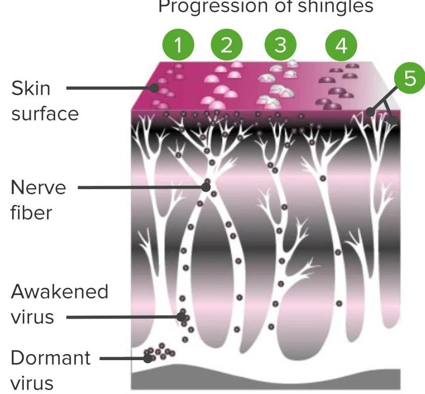
Herpes Zoster (shingles) presents as a blistering rash following a specific dermatomal distribution, most commonly in the thoracic region. While it may occasionally cross the midline (about 20% of cases), it is typically localized to a nerve pathway.
Diagram illustrating the progression of shingles, showing the skin surface, nerve fibers, and the awakening and dormancy of the virus.
A significant complication is post-herpetic neuralgia (PHN), which affects approximately 13% of individuals over age 60.
Management of Herpes Zoster:
Antiviral Agents: These can reduce the duration of symptoms and viral shedding if administered within 72 hours of rash onset.
Prevention: The "shingles" vaccine, recommended at age 60, is highly effective at preventing both the disease and PHN.
Generalized Rashes and Immune-Mediated Drug Reactions
Generalized rashes involve larger body surfaces and often suggest systemic or immune-mediated processes.
Viral Exanthems
Fifth Disease: Caused by parvovirus B19, it presents with a "slapped cheek" appearance and a subsequent fishnet-pattern rash.
Roseola: Typically seen in children under 3, it begins with a high fever; once the fever subsides, a maculopapular rash spreads from the center to the periphery.
Psoriasis and Pityriasis Rosea
Psoriasis affects about 2% of U.S. adults, with 90% presenting in the plaque form, often on extensor surfaces. Management depends on the extent of involvement:
< 5% involvement: Topical corticosteroids or calcineurin inhibitors.
> 5% involvement or arthritis: Systemic treatments like phototherapy or biologic agents (e.g., TNF inhibitors).
Pityriasis Rosea presents as oval, salmon-colored patches in a "Christmas tree" pattern on the back, often preceded by a prominent "herald patch". It is generally self-limiting and requires only reassurance.
Acute Cutaneous Drug Reactions
Drug reactions are categorized by their underlying mechanisms.
1. Exanthematous Reactions
This is the most common type of cutaneous drug reaction (90%). It presents as a symmetric, pruritic, bright red maculopapular eruption on the trunk and extremities. Common culprits include antibiotics (sulfas, beta-lactams), anticonvulsants, and NSAIDs.
2. Urticaria and Angioedema
Urticaria (hives) involves mast cell activation in the superficial dermis, causing intensely pruritic, well-demarcated wheals. Triggers include beta-lactam antibiotics, insect bites, and infections.
Angioedema is the localized edema of deeper soft tissues or mucosal tissues (lips, face, tongue, throat). It can be mast cell-mediated or bradykinin-mediated (often due to ACE inhibitors).
Warning: Angioedema can be life-threatening if it involves the throat, causing airway obstruction.
3. Severe Cutaneous Adverse Reactions (SCARs)
These are high-morbidity conditions that require immediate intervention:
Erythema Multiforme (EM): Characterized by targetoid lesions (circular, target-like) distributed symmetrically on the hands and face. It is often triggered by infections like HSV.
Stevens-Johnson Syndrome (SJS) and Toxic Epidermal Necrolysis (TEN): These involve a T-cell-mediated attack on keratinocytes. SJS involves < 10% of the skin, while TEN involves > 30%. Both present with fever, mucosal involvement (lips, eyes, genitalia), and a positive Nikolsky sign.
DRESS/DHS: A delayed hypersensitivity reaction (2–8 weeks after drug exposure) involving eosinophilia and multi-organ involvement (e.g., liver, kidneys).
Management of SJS/TEN:
Discontinue the culprit drug immediately.
Admit to an ICU or burn unit.
Provide intensive supportive care (IV fluids, wound care, nutrition).
Exam Tip: When evaluating a patient with a rash, always check for mucosal involvement and the Nikolsky sign. The presence of both, along with systemic symptoms, strongly points toward SJS/TEN rather than Erythema Multiforme.
Keep reading for free.
Create an account to finish this reading and make it part of your study routine.