Lipids, Nucleic Acids, and Proteins: The Molecular Building Blocks of Life
An overview of the three major classes of biological macromolecules: lipids, nucleic acids, and proteins. Explores their structures, functions, and roles in cellular processes, including energy storage, genetic information, and cellular machinery. Discusses protein folding, misfolding, and related diseases.
Life is governed by a complex interplay of specialized molecules that perform distinct roles, from storing genetic blueprints to driving metabolic reactions. To understand the molecular mechanisms of disease, one must first master the three major classes of biological macromolecules: lipids, nucleic acids, and proteins. While each class possesses unique chemical properties, they collectively provide the structure, information, and machinery required for cellular survival.
Lipids: Energy, Membranes, and Signaling
Lipids are a diverse group of molecules characterized by their hydrophobicity (water-fearing) or their amphipathic nature, meaning they contain both hydrophilic (water-loving) and hydrophobic regions. Unlike proteins or nucleic acids, lipids are not polymers made of repeating monomeric units, but they are categorized into major classes based on their chemical structures.
Fats (Triglycerides) and Fatty Acids
Fats, or triglycerides, are highly efficient molecules for long-term energy storage. They consist of a single glycerol backbone linked to three fatty acids via ester bonds.
A fatty acid is composed of a carboxyl group ($-COOH$) and a long hydrocarbon chain. The physical properties of these fats depend heavily on the saturation of the fatty acid chains:
Saturated fatty acids: These contain no double bonds in the hydrocarbon chain. This allows the chains to be straight, enabling them to pack tightly together.
Unsaturated fatty acids: These contain one or more double bonds. In a cis orientation, these double bonds create "kinks" in the chain, preventing tight packing.
Phospholipids and the Lipid Bilayer
Phospholipids are the fundamental building blocks of cell membranes. They are amphipathic, consisting of a glycerol backbone, two hydrophobic fatty acid tails, and a phosphate-containing polar head that is hydrophilic.
When placed in an aqueous environment, these molecules spontaneously assemble into a lipid bilayer. The hydrophobic tails cluster together to avoid water, while the hydrophilic heads face the water on both sides of the membrane. This assembly is driven by the hydrophobic effect rather than covalent bonding between individual lipids.
Diagram illustrating the amphipathic structure of a molecule, showing a polar phosphate head that interacts with water and hydrophobic fatty acid tails that avoid water.
Steroids and Cholesterol
Steroids represent a class of lipids with a structure fundamentally different from fats or phospholipids. They are built upon a characteristic four-ring hydrocarbon skeleton.
Cholesterol is perhaps the most vital steroid in animal cells. It serves two primary roles:
Membrane Component: It is integrated into the lipid bilayer to help regulate membrane fluidity.
Precursor Molecule: It serves as the starting material for the synthesis of steroid hormones (such as estrogen, testosterone, and cortisol), Vitamin D, and bile acids.
Nucleic Acids: The Information Carriers
Nucleic acids are polymers of nucleotides that serve as the primary means of storing and transmitting genetic information.
The Structure of a Nucleotide
Every nucleotide is composed of three essential components:
A five-carbon sugar (ribose in RNA or deoxyribose in DNA).
A phosphate group.
A nitrogenous base (Adenine, Guanine, Cytosine, and either Thymine or Uracil).
The distinction between the two main types of nucleic acids lies in their sugar and their bases:
DNA (Deoxyribonucleic acid): Uses deoxyribose sugar (which has a hydrogen atom at the 2' position, $2'–H$) and the base Thymine (T). It is responsible for long-term genetic storage.
RNA (Ribonucleic acid): Uses ribose sugar (which has a hydroxyl group at the 2' position, $2'–OH$) and the base Uracil (U). It is primarily involved in gene expression and protein synthesis.
Nucleotides are linked together by 3'–5' phosphodiester bonds, which connect the 3'–OH group of one sugar to the 5' phosphate of the next, creating a continuous sugar-phosphate backbone.
Diagram illustrating the molecular structure of a nucleotide, composed of a phosphate group, a pentose sugar, and a nitrogenous base.
Beyond Genetics: The Versatility of Nucleotides
Nucleotides are not limited to storing information; they are also essential for cellular metabolism and signaling:
ATP (Adenosine triphosphate): The universal energy currency of the cell.
GTP: Provides energy for specific reactions and powers G protein signaling.
cAMP (Cyclic AMP): Acts as a vital intracellular messenger.
Coenzyme A (CoA): Carries acyl groups (like acetyl groups) during metabolic processes.
Proteins: The Cellular Machinery
Proteins are large macromolecules that act as the "molecular tools and machines" of the cell. They perform nearly all cellular activities, including catalysis (enzymes), transport, signaling (hormones), and structural support.
Amino Acids and Polypeptide Structure
The building blocks of proteins are amino acids. Each amino acid features a central α-carbon, an amino group (−NH3+), a carboxyl group ($-COO^-$), and a variable R group (side chain) that determines its chemical properties.
Amino acids are categorized by their R groups:
Polar charged: Can form ionic bonds (e.g., Lysine, Glutamate).
Polar uncharged: Can form hydrogen bonds (e.g., Serine).
Nonpolar: Hydrophobic and help stabilize the protein core (e.g., Leucine).
Special cases: Cysteine is unique because its sulfhydryl ($-SH$) group can form covalent disulfide bridges, which are critical for stabilizing protein structure.
The Four Levels of Protein Organization
A protein's function is entirely dependent on its unique three-dimensional shape. This shape is organized into four levels:
Primary structure: The linear sequence of amino acids.
Secondary structure: Local conformations like α-helices and β-sheets, stabilized by backbone hydrogen bonds.
Tertiary structure: The full 3D conformation of a single polypeptide, stabilized by R-group interactions.
Quaternary structure: The arrangement of multiple polypeptide subunits (e.g., the four subunits of hemoglobin).
Exam Tip: Remember that denaturation is the process where a protein loses its 3D structure (and thus its function) due to external stressors like heat, radiation, or detergents, even though the primary sequence remains intact.
Protein Misfolding and Disease
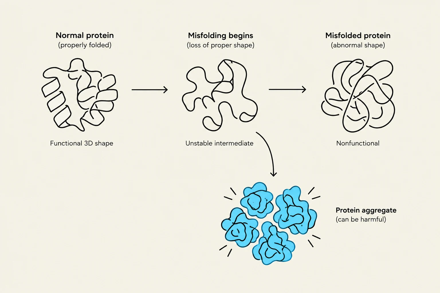
While most proteins fold into low-energy states, some can misfold into abnormal shapes. Misfolded proteins can expose hydrophobic surfaces that cause them to aggregate into stable, toxic structures called amyloid fibrils.
A notable example is the prion protein (PrP). In its normal state ($PrP^C$), it is functional, but it can transition into a misfolded, infectious state (PrPSc). This misfolded protein can "template" or induce normal proteins to also misfold, leading to neurodegenerative diseases such as Creutzfeldt–Jakob disease (CJD).
To combat misfolding, cells utilize molecular chaperones (helper proteins). The Hsp70 family shields emerging polypeptides, while chaperonins provide isolated chambers to help complex proteins fold correctly.
This diagram illustrates the process of protein misfolding, where a normally folded protein loses its proper shape, becoming nonfunctional and potentially forming harmful aggregates.
Keep reading for free.
Create an account to finish this reading and make it part of your study routine.