Explores the molecular structure of lipids, the properties of phospholipid bilayers, and the mechanisms of cellular transport including diffusion, osmosis, and active transport.
The cell membrane is more than just a simple container; it is a sophisticated, dynamic barrier that separates life from nonlife. By acting as a selective barrier, the membrane controls which molecules enter and exit the cell, while also helping to concentrate reactants to speed up essential chemical reactions. To understand how cells maintain this delicate balance, we must first look at the molecular building blocks that make up the membrane: lipids.
The Molecular Structure of Lipids
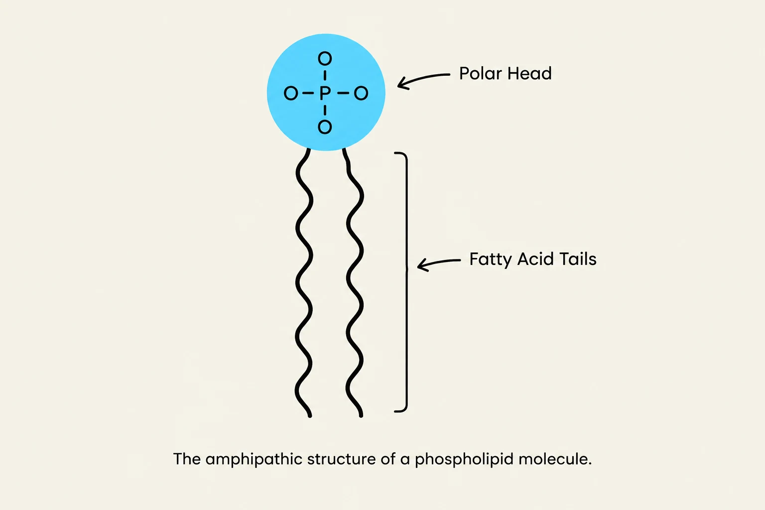
The amphipathic structure of a phospholipid molecule, showing its polar head and fatty acid tails.
Lipids are a diverse group of molecules defined primarily by their hydrophobic nature, meaning they do not dissolve in water. This property is largely due to the presence of many nonpolar C-H (carbon-hydrogen) bonds. While they share this common trait, lipids fall into several distinct chemical classes:
Triacylglycerols (Fats): These consist of a single glycerol molecule linked to three fatty acids via ester linkages. Because the fatty acids are not linked in a continuous chain, fats are not considered polymers.
Steroids: These molecules feature a characteristic nonpolar four-ring structure. A well-known example is cholesterol, which plays a vital role in membrane stability.
Phospholipids: These are the fundamental components of cell membranes.
Phospholipids are unique because they are amphipathic, possessing both a polar "hydrophilic" (water-loving) head and a nonpolar "hydrophobic" (water-fearing) tail. The head consists of a three-carbon glycerol attached to a phosphate group (PO4−), while the tails are composed of two fatty acids.
The Phospholipid Bilayer and the Fluid-Mosaic Model
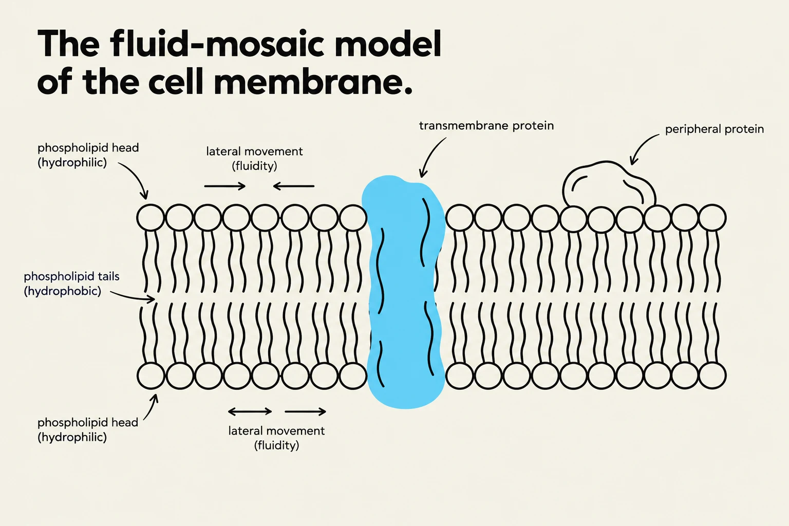
Diagram illustrating the fluid-mosaic model of a cell membrane, showing phospholipid heads and tails, transmembrane and peripheral proteins, and lateral movement.
When placed in water, phospholipids spontaneously organize themselves to minimize contact between the hydrophobic tails and the aqueous environment. This results in two common structures: micelles (tiny droplets with heads outside and tails inside) or lipid bilayers. In a bilayer, two sheets of phospholipids sit back-to-back, with the nonpolar tails facing inward toward each other and the polar heads facing the water on both sides.
The modern understanding of this structure is the fluid-mosaic model. This model describes the membrane as a dynamic, fluid environment where phospholipids move laterally (side-to-side) within the bilayer. Embedded within this fluid sea of lipids are various proteins:
Transmembrane proteins span the entire membrane.
Peripheral membrane proteins are attached to only one side of the membrane.
Exam Tip: Remember that membrane proteins are also amphipathic. Their nonpolar amino acids are tucked into the hydrophobic interior of the bilayer, while their polar amino acids "stick out" into the aqueous environment on either side.
Factors Affecting Membrane Fluidity and Permeability
The membrane must maintain a specific level of fluidity to function. Several factors influence how "liquid" or "solid" the membrane is:
Saturation of Fatty Acid Tails:Saturated lipids have straight tails with no double bonds, allowing them to pack tightly together. This increases membrane density but reduces both fluidity and permeability. Unsaturated lipids contain C=C double bonds that create "kinks" in the tails. These kinks prevent tight packing, thereby increasing fluidity and permeability.
Cholesterol: This steroid molecule dramatically reduces membrane permeability.
Temperature: As temperature drops, hydrophobic tails pack more closely together. This decreases fluidity and can cause the membrane to solidify if the temperature becomes low enough.
Selective Permeability
The lipid bilayer is selectively permeable, meaning it allows some substances to pass through more easily than others.
High Permeability: Small, nonpolar molecules (like O2) can cross the membrane very quickly.
Low Permeability: Large molecules (like glucose or sucrose) and charged ions (like $K^+$ or $Cl^-$) cross very slowly, if at all, without the help of proteins.
Mechanisms of Cellular Transport
Comparison of active and passive transport mechanisms.
To move substances that cannot cross the bilayer alone, cells utilize various transport mechanisms. These are categorized based on whether they require energy.
Passive Transport: Diffusion and Osmosis
Passive transport is powered solely by the movement of molecules along an electrochemical gradient (the combined effect of concentration and electrical charge). It does not require an input of cellular energy.
Diffusion is the spontaneous movement of solutes from an area of high concentration to an area of low concentration. This process is driven by the thermal energy and constant motion of molecules, which increases entropy. When molecules are distributed equally throughout a space, the system has reached equilibrium.
Osmosis is a specific type of diffusion referring to the movement of water. Water moves from areas of low solute concentration to areas of high solute concentration. The effect of osmosis depends on the surrounding solution:
Isotonic: The solute concentration is equal inside and outside the cell; there is no net movement of water.
Hypertonic: The solute concentration is higher outside the cell. Water moves out of the cell, causing it to shrink.
Hypotonic: The solute concentration is lower outside the cell. Water moves into the cell, causing it to swell and potentially burst.
Facilitated Diffusion
Some substances require help to move down their concentration gradient via facilitated diffusion. This is achieved through membrane proteins:
Channel Proteins: These form selective pores. Some are gated, meaning they open or close in response to specific signals. For example, aquaporins are specialized channels for water.
Carrier Proteins: Unlike channels, these do not form a continuous pore; instead, they change their shape to move a substance across the membrane.
Active Transport
Active transport moves molecules against their concentration gradient (from low to high concentration). Because this movement is "uphill," it requires energy, typically in the form of ATP.
A classic example is the sodium-potassium pump. For every single ATP molecule used, the pump transports three Na+ ions out of the cell and two $K^+$ ions into the cell.
Finally, cells use secondary active transport (also known as cotransport). In this process, a protein pump first uses ATP to establish an electrochemical gradient. The cell then uses the potential energy stored in that gradient to power the transport of a different molecule against its own gradient.
Summary Table: Transport Comparison
Feature
Passive Transport
Active Transport
Direction
High → Low concentration
Low → High concentration
Energy (ATP) Required?
No
Yes
Driving Force
Electrochemical gradient
ATP or established gradients
Examples
Diffusion, Osmosis, Facilitated Diffusion
Sodium-potassium pump, Cotransport
By coordinating these lipids and proteins, the cell creates an internal environment that is chemically distinct from the outside world, allowing life to persist.
Keep reading for free.
Create an account to finish this reading and make it part of your study routine.