Leukocytes and Platelets: The Body's Defense and Repair Systems
An overview of leukocytes (white blood cells) and platelets, detailing their classification, functions in immunity and hemostasis, and their production through hematopoiesis. Includes a discussion on erythropoiesis and the regulation of hematocrit.
While red blood cells are the most numerous components of blood, they are not the only essential formed elements. Blood is a complex fluid connective tissue composed of plasma and formed elements, including erythrocytes (red blood cells), leukocytes (white blood cells), and platelets. While erythrocytes focus on gas exchange, leukocytes and platelets serve critical roles in immunity and hemostasis.
Leukocytes: The Immune Sentinels
Leukocytes are larger than erythrocytes and possess prominent nuclei. Interestingly, unlike red blood cells which function primarily within the circulatory system, leukocytes typically use the blood as a vehicle to reach various tissues throughout the body where they perform their specific immune functions.
Leukocytes are classified into two main categories based on the presence or absence of visible cytoplasmic granules: granulocytes and agranulocytes.
Granulocytes
Granulocytes are characterized by visible cytoplasmic granules and unusually shaped, multi-lobed nuclei. This group includes three distinct cell types:
Neutrophils: These are the most abundant leukocytes, comprising 60-70% of the total population. They are polymorphonuclear cells, meaning they possess 3 to 5 variable nuclear lobes. Neutrophils are highly phagocytic and specialize in destroying bacterial cells. When they exit the bloodstream to reach an infection site, they release granules that kill bacteria, enhance inflammation, and attract additional leukocytes to the area.
Eosinophils: Making up approximately 4% of leukocytes, these cells feature a bilobed nucleus and take up acidic red eosin dye. They act as phagocytes that respond specifically to parasitic worm infections and allergic reactions.
Basophils: These are the rarest leukocytes, representing less than 1% of the population. They take up basic methylene blue dye, and their numerous granules are often so dense that they obscure the cell's S-shaped nucleus. Basophils release chemicals that mediate inflammation.
Diagram illustrating three types of granulocytes: Neutrophil, Eosinophil, and Basophil, detailing their nucleus shapes and functions.
Agranulocytes
Agranulocytes lack visible cytoplasmic granules and consist of two main types:
Lymphocytes: Accounting for 20-25% of leukocytes, these cells are characterized by a large, spherical nucleus surrounded by a thin rim of cytoplasm. They are divided into two functional groups: B lymphocytes, which mature in the bone marrow and produce antibodies, and T lymphocytes, which mature in the thymus gland and activate other components of the immune system.
Monocytes: These are the largest leukocytes, making up 3-8% of the population. They possess a large, U-shaped nucleus. When monocytes exit the capillaries and enter body tissues, they mature into macrophages. Macrophages are powerful phagocytes that ingest bacteria, antigens, dead or dying cells, and other cellular debris. Furthermore, they activate the broader immune system by displaying phagocytosed antigens to other leukocytes.
Exam Tip: Differentiation
Remember that the distinction between B and T lymphocytes is their site of maturation: B cells in the Bone marrow, and T cells in the Thymus.
Platelets: The First Responders to Injury
Platelets are the smallest of the formed elements and are essential for hemostasis, the process of blood clotting. Unlike the larger leukocytes, platelets are actually cell fragments that possess a plasma membrane but lack nuclei and most organelles. They contain granules filled with clotting factors and enzymes, as well as cytoskeletal elements and microtubules to facilitate their function.
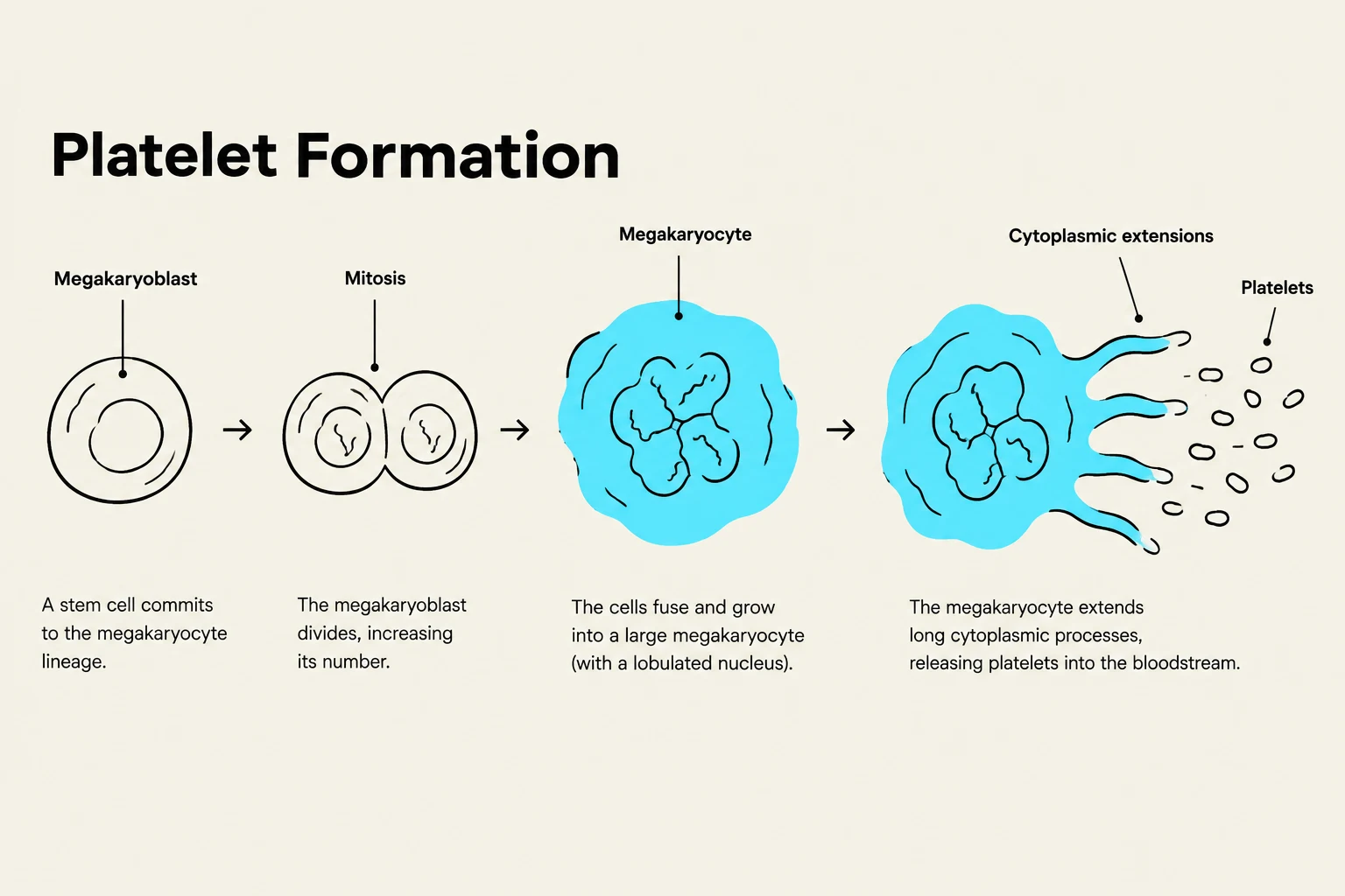
Platelet formation (thrombopoiesis) begins with hematopoietic stem cells in the red bone marrow. They develop through the myeloid cell line, progressing from a megakaryoblast to a large megakaryocyte. A megakaryocyte undergoes repeated rounds of mitosis without cytokinesis, resulting in a massive cell with multiple copies of DNA. These cells mature in response to the hormone thrombopoietin and extend ribbon-like arms into the bone marrow sinusoids. The force of blood flow then breaks off these arms, creating newly formed platelets.
Diagram illustrating the process of platelet formation, starting from a stem cell committing to the megakaryocyte lineage, followed by mitosis, growth into a large megakaryocyte, and finally the release of platelets through cytoplasmic extensions.
Hematopoiesis and Regulation
All formed elements, including leukocytes, erythrocytes, and platelets, are produced in the red bone marrow through a process called hematopoiesis. This process begins with hematopoietic stem cells (HSCs), which can differentiate into two distinct lines: the myeloid cell line (producing monocytes, granulocytes, erythrocytes, and platelets) and the lymphoid cell line (producing lymphocytes).
Erythropoiesis: A Specialized Example
While leukocytes are produced via leukopoiesis, the production of erythrocytes is specifically called erythropoiesis. Because erythrocytes have a relatively short lifespan of only 100-120 days, continuous production is vital. The developmental stages include:
Progenitor Erythrocyte Colony Forming Units (CFUs): Cells committed to the erythrocyte pathway.
Proerythroblasts: Formed from CFUs; their development is driven by the hormone erythropoietin (EPO).
Erythroblasts: These cells synthesize hemoglobin and other proteins. During the late stage, the nucleus shrinks and is eventually ejected.
Reticulocytes: These are nucleus-free cells that are in the final stages of ejecting remaining organelles. They enter the bloodstream through the sinusoidal capillaries of the bone marrow, a process taking about 5-7 days.
Erythrocytes: The final, mature, anucleated red blood cell.
The Negative Feedback Loop
The body maintains an optimal hematocrit (the percentage of blood volume occupied by erythrocytes) through a negative feedback loop. If the hematocrit is too low, oxygen transport to tissues becomes insufficient; if it is too high, the blood becomes too viscous, leading to sluggish flow.
The primary regulator is erythropoietin (EPO), a hormone secreted by the kidneys. When the kidneys detect low oxygen (O2) levels in the blood, they increase the secretion of EPO. This hormone stimulates the bone marrow to increase the rate of erythropoiesis and accelerate erythrocyte maturation, which ultimately raises the hematocrit back to a homeostatic range.
Summary Table: Leukocyte Classification
Type
Category
Key Feature
Primary Function
Neutrophil
Granulocyte
3-5 nuclear lobes
Phagocytosis of bacteria
Eosinophil
Granulocyte
Bilobed nucleus
Parasites & Allergies
Basophil
Granulocyte
S-shaped nucleus
Inflammation
Lymphocyte
Agranulocyte
Large spherical nucleus
Adaptive immunity (B & T cells)
Monocyte
Agranulocyte
U-shaped nucleus
Becomes Macrophage (Phagocytosis)
Keep reading for free.
Create an account to finish this reading and make it part of your study routine.