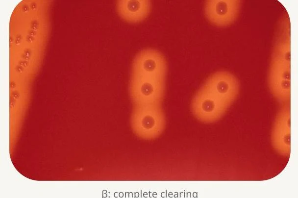
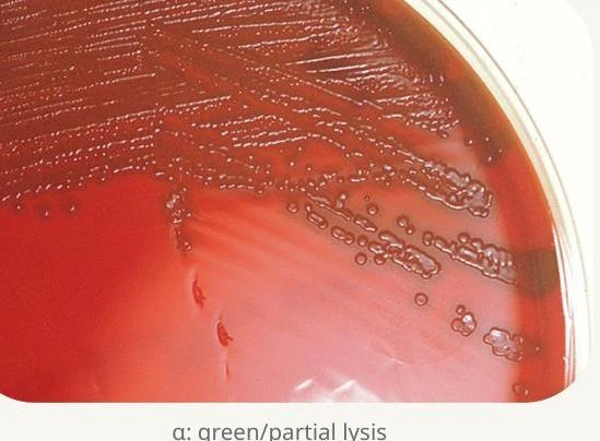
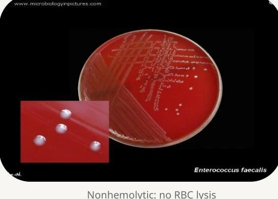
β-Hemolytic Streptococci
β-hemolytic organisms are often identified by their Lancefield group antigens, which are carbohydrates located in the bacterial cell wall.
Group A Streptococcus (S. pyogenes)
S. pyogenes is a major human pathogen responsible for pharyngitis (strep throat), scarlet fever, and skin infections like impetigo and erysipelas. It can also cause life-threatening conditions such as necrotizing fasciitis and toxic shock syndrome.
To confirm S. pyogenes, laboratories look for:
- Bacitracin susceptibility (TAXO-A): A zone of inhibition around the bacitracin disk indicates susceptibility.
- PYR test: A positive result shows a red or pink color change.
Group B Streptococcus (S. agalactiae)
S. agalactiae is a significant concern in obstetrics, as it is a major cause of neonatal sepsis and meningitis. Pregnant women are routinely screened via vaginal and rectal swabs.
Identification is confirmed through:
- CAMP test: This test looks for an "arrowhead-shaped" zone of enhanced hemolysis where the organism meets Staphylococcus aureus.
- Sodium Hippurate Hydrolysis: A positive result turns the medium purple after the addition of ninhydrin.
Exam Tip: If you see a β-hemolytic isolate from a pregnancy screen that is CAMP positive and Hippurate positive, think Group B Streptococcus (S. agalactiae).
Groups C and G
These groups can clinically resemble Group A Streptococcus (GAS) and may cause similar pyogenic infections. Because their morphology overlaps with GAS, Lancefield typing is the gold standard for definitive differentiation.
α-Hemolytic Streptococci
The α-hemolytic group is primarily divided into S. pneumoniae and the Viridans group.
Streptococcus pneumoniae (Pneumococcus)
S. pneumoniae is a lancet-shaped diplococcus and a leading cause of pneumonia, meningitis, and otitis media. Its virulence is heavily driven by its polysaccharide capsule.
The laboratory distinguishes pneumococcus from other α-strep using:
- Optochin susceptibility (TAXO-P): A zone of inhibition ≥14 mm indicates S. pneumoniae.
- Bile solubility: The addition of sodium deoxycholate causes the colonies to disappear or clear.
Viridans Streptococci
These are normal flora of the oral cavity and gastrointestinal tract. While generally low in virulence, they can cause opportunistic infections such as subacute bacterial endocarditis. Unlike pneumococcus, Viridans streptococci are Optochin resistant and Bile insoluble.
γ-Hemolytic (Non-hemolytic) Streptococci
Non-hemolytic organisms are often identified by their ability to grow in harsh environments and their reaction to bile and salt.
Enterococcus spp.
Formerly classified as Group D streptococci, Enterococcus species are common nosocomial (hospital-acquired) pathogens, frequently causing UTIs and bacteremia. They are distinguished by:
- Bile esculin hydrolysis: A positive result produces a black precipitate.
- 6.5% NaCl tolerance: They can grow in high salt concentrations, resulting in broth turbidity.
- PYR positivity: A positive result shows a red/pink color change.
Group D Streptococci (S. gallolyticus group)
This group is distinct from Enterococcus because they are 6.5% NaCl intolerant and PYR negative. Clinically, this group is highly significant due to its strong association with colorectal disease and endocarditis.
Advanced Diagnostic Methodologies
While traditional culture on blood agar remains the cornerstone of diagnosis—especially for performing antimicrobial susceptibility testing—modern laboratories utilize several rapid methods:
- Immunoassays: These detect group-specific antigens directly from colonies or swabs.
- NAAT/PCR: These molecular methods detect specific bacterial genes and provide very rapid results.
- MALDI-TOF: This advanced technology allows for rapid identification of organisms directly from isolated colonies.
Antimicrobial Susceptibility Testing
Susceptibility testing is critical for managing emerging resistance patterns. Clinicians must be particularly vigilant regarding Vancomycin-resistant Enterococcus (VRE) and increasing penicillin resistance in S. pneumoniae.
Summary Table for Rapid Identification
| Organism | Hemolysis | Key Confirmatory Test |
|---|
| S. pyogenes (GAS) | β | Bacitracin Susceptible / PYR (+) |
| S. agalactiae (GBS) | β | CAMP (+) / Hippurate (+) |
| S. pneumoniae | α | Optochin Susceptible / Bile Soluble |
| Enterococcus spp. | α,β,γ | 6.5% NaCl (+) / PYR (+) |
| S. gallolyticus (Group D) | α,γ | 6.5% NaCl (-) / PYR (-) |