Joints and Articular Cartilage: Structure, Function, and Types
Explore the anatomy and classification of joints, including synarthroses (fibrous and cartilaginous) and diarthroses (synovial). Learn about the structure of synovial joints, the mechanics of joint movement (uniaxial, biaxial, triaxial), and the composition and function of articular cartilage. This content also covers
In the human body, a joint (or articulation) is defined as the point of contact between two bones. The specific configuration of a joint is the primary factor that dictates its possible range of movement. Understanding joints requires a dual focus: first, the anatomical classification based on how the bones are held together, and second, the mechanical function of the tissues that allow for smooth, frictionless motion.
Classification of Joints: Synarthroses vs. Diarthrosis
Joints are broadly categorized by their degree of mobility. The first major category is synarthroses, which are joints designed to allow little or no movement. These are subdivided into two types:
Fibrous Joints: Bones are united by dense fibrous tissue.
Suture joints: Found only in the skull, where bones are united by a thin, dense layer of fibrous tissue.
Gomphosis joints: A "peg-in-a-hole" configuration, specifically the joint between the teeth and the mandible.
Syndesmosis joints: Two bones joined by a ligament or an interosseous membrane, such as the connection between the tibia and fibula.
Cartilaginous Joints: Bony segments are connected by either fibrocartilage or hyaline growth cartilage.
Symphysis joints: These connect bony segments via a fibrocartilaginous plate or disk, such as the symphysis pubis.
Synchondrosis joints: These involve cartilage connecting bony components, often found at growth sites in the body, such as the first sternocostal joint.
Diagrams illustrating fibrous and cartilaginous joints in the human body, including sutures, gomphosis, syndesmosis, symphysis, and costochondral joints, with key differences highlighted.
In contrast to the stability of synarthroses, diarthrosis (also known as synovial joints) are designed specifically for mobility.
The Anatomy of Synovial Joints
Synovial joints are complex structures that facilitate movement through a coordinated system of tissues. A diarthrosis is enclosed by a joint capsule or synovial sheath. The internal structure includes:
Joint Cavity: The space enclosed by the capsule.
Synovial Membrane: The inner surface of the capsule.
Synovial Fluid: A lubricating fluid produced by fibroblast-like cells lining the joint capsule. This fluid coats the surfaces to reduce friction.
Articular Cartilage: The smooth tissue covering the ends of the bones.
Clinical Note: Movement in these joints is provided by the contraction of the muscle-tendon unit, while the control and stability of that movement depend heavily on the joint capsule and surrounding ligaments.
Diagram illustrating the anatomy of a synovial joint, including bones, articular cartilage, joint capsule, synovial membrane, and synovial fluid.
Mechanics of Joint Movement
The way a joint moves is categorized by the number of axes it can rotate around. These movements include flexion, extension, rotation, adduction, and abduction.
Uniaxial Joints
These allow motion around a single axis:
Hinge joints: Permit flexion and extension (e.g., elbow, knee, or interphalangeal joints of the fingers).
Pivot joints: Allow rotation around a single axis (e.g., the superior radioulnar joint of the elbow).
Biaxial Joints
These permit movement in two planes:
Condyloid joints: Allow flexion and extension at one axis and adduction and abduction at another (e.g., metacarpophalangeal joints of the hand).
Saddle joints: Characterized by surfaces that are convex in one plane and concave in another (e.g., the carpometacarpal joint of the thumb).
Triaxial Joints
These permit movement around three axes, allowing motion in three planes:
Carpal joints: Involve gliding or rotating relative to adjacent surfaces.
Ball-and-socket joints: Formed by a ball-like surface fitting into a concave socket, providing maximum mobility (e.g., hip and shoulder).
Articular Cartilage: Structure and Function
Articular cartilage is a specialized tissue that appears smooth, shiny, and white. Its primary role is to cover the ends of bones, distribute joint loads over a wide area, and allow for movement with minimal friction.
Composition and Properties
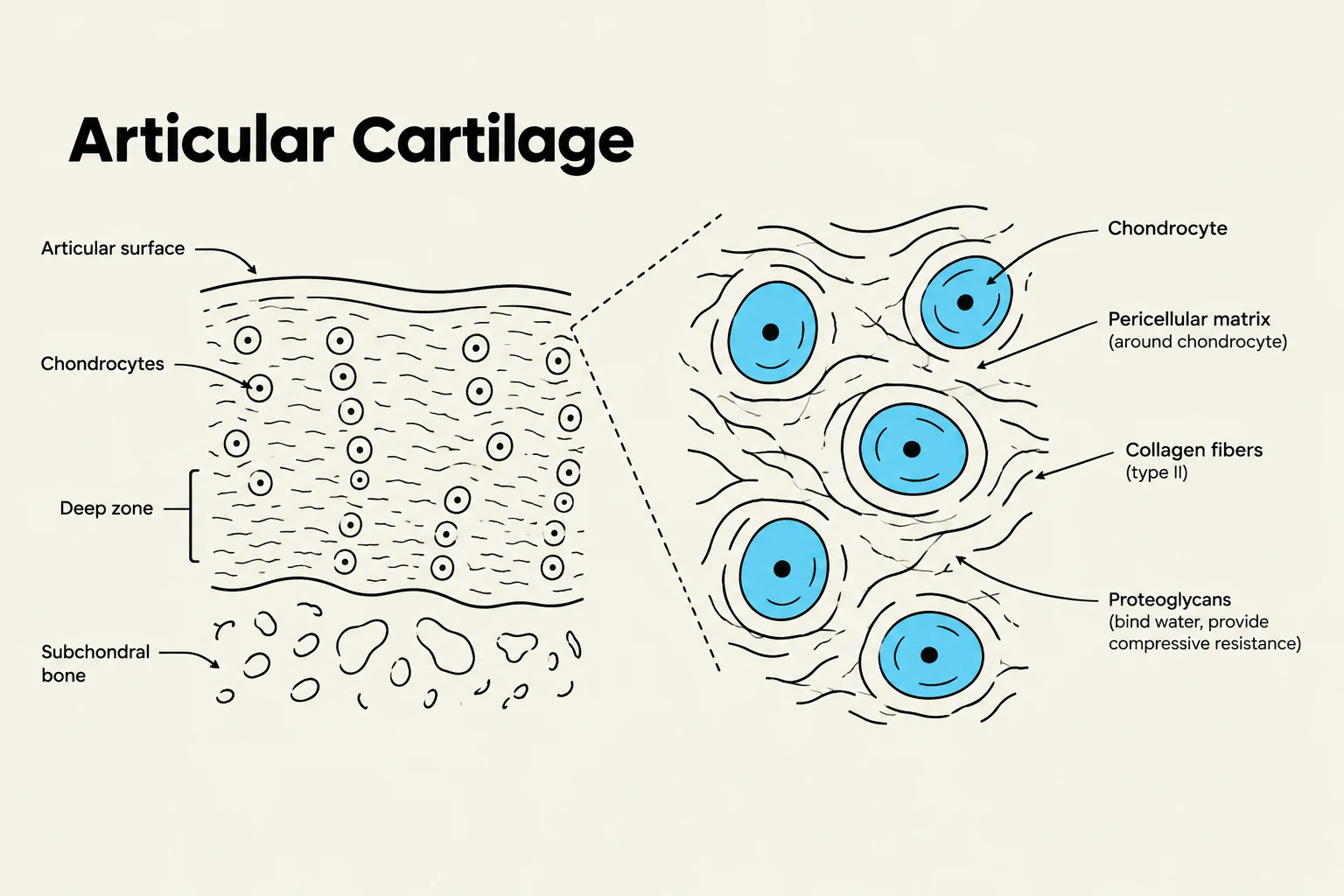
The cartilage is primarily composed of water, inorganic salts, proteins, glycoproteins, and lipids. The extracellular matrix is made of collagen fibers (elastin and collagen) and a network of collagen fibrils encased in a solution of proteoglycans, which are manufactured by cells called chondrocytes.
The functional properties of this tissue are highly specialized:
Collagen fibers: Provide stability, strength, and tensile stiffness; they are excellent at tolerating tension but not compression.
Proteoglycan aggregation: Provides structural rigidity and allows the tissue to tolerate compression.
Load Distribution: By spreading applied loads over a large area, the cartilage decreases contact stress and limits wear.
Exam Tip: Remember that articular cartilage is devoid of blood vessels, lymph channels, and nerves. This lack of vascularity is a critical reason why cartilage has a limited capacity for self-repair.
Diagram illustrating the structure of articular cartilage, including chondrocytes, pericellular matrix, collagen fibers, proteoglycans, and subchondral bone.
Clinical Considerations: Injury and Degeneration
Because articular cartilage relies on lubrication and structural integrity, it is susceptible to several types of damage:
Interfacial wear: Caused by insufficient lubrication.
Fatigue wear: Resulting from repetitive stress injuries.
Trauma: Sudden imposition of excessive stress can damage the joint matrix.
Degeneration: Over time, wear can cause microcracks and the formation of "loose bodies." This results in a rough, irregular surface that can lead to osteoarthritis or degenerative joint disease.
Furthermore, clinicians must distinguish between different types of joint trauma. A dislocation involves the complete separation of the articulating surfaces, whereas a subluxation refers to only a partial separation. Both conditions can cause significant tissue damage, pain, changes in the joint's contour, and loss of mobility.
Keep reading for free.
Create an account to finish this reading and make it part of your study routine.