Veterinary dentistry is a specialized area of veterinary medicine dedicated to maintaining oral health, managing teeth, and preventing or treating dental diseases. For veterinary technicians, understanding the intricate anatomy of the mouth is not just an academic exercise; it is a clinical necessity. Mastery of these structures allows for accurate oral charting, effective dental radiography, and the ability to recognize subtle abnormalities during examinations that might otherwise be missed.
Skeletal Framework and Head Shapes
The foundation of the oral cavity is provided by the skull and jawbones. The maxilla forms the upper jaw, while the mandible forms the lower jaw. These two structures are connected by the temporomandibular joint (TMJ), which allows the mandible to move, enabling the animal to open and close its mouth.
When examining patients, it is vital to consider their cranial morphology, as head shape can significantly impact dental health:
Brachycephalic: These animals have short muzzles and wide heads (e.g., Bulldogs, Pugs, and Persian cats). Due to the shortened muzzle, premolars are often crowded or rotated.
Mesocephalic: This is the most common head shape, characterized by a medium length and width muzzle (e.g., Golden Retrievers and domestic shorthair cats).
Dolichocephalic: These animals possess long, narrow heads and muzzles (e.g., Greyhounds and Siamese cats).
Clinical Tip: When performing a dental exam on a brachycephalic patient, always document tooth crowding and abnormal positioning, as these are common findings in this head shape.
Detailed Tooth Structure
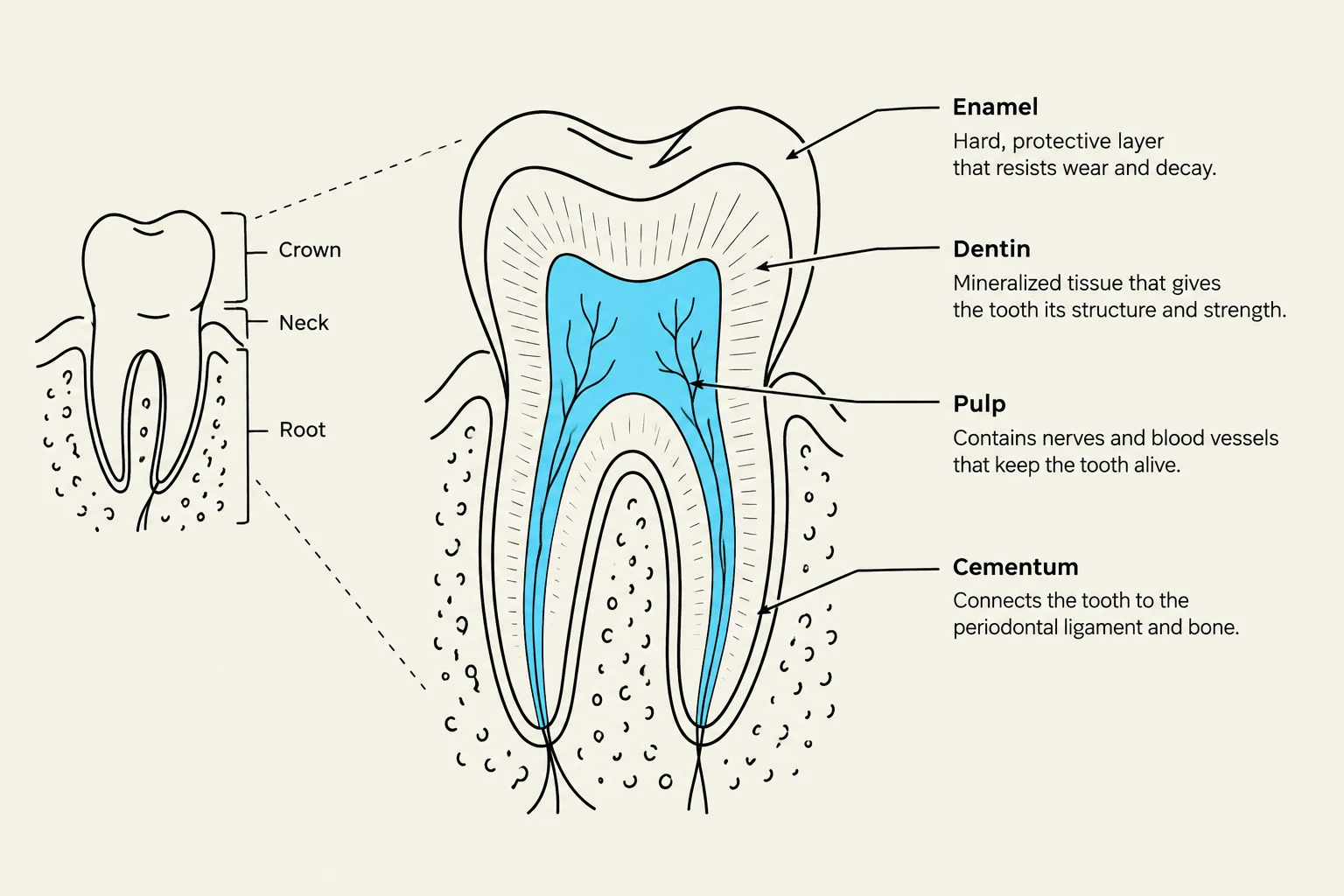
A tooth is a complex organ composed of several distinct layers, each with a specific biological function. A tooth is divided into two main parts: the crown, which is the visible portion above the gumline, and the root, which is embedded within the jawbone.
Internal and External Layers
The various tissues that make up a tooth include:
Enamel: The hardest tissue in mammals, enamel forms the outer covering of the tooth crown. It develops before eruption and gradually wears down over the animal's lifetime.
Dentin: This tissue makes up the bulk of the tooth and surrounds the pulp. It is harder than bone but softer than enamel, and it is capable of detecting touch, heat, and cold. Specialized cells called odontoblasts are responsible for producing dentin.
Pulp: The innermost core of the tooth, containing nerves, blood vessels, and connective tissue. Because it is highly vascularized and innervated, the pulp is a critical consideration when evaluating dental fractures.
Cementum: A hard, calcified tissue that covers the root dentin, helping to support the tooth and assist in root repair.
Diagram illustrating the different parts of a tooth: enamel, dentin, pulp, and cementum, along with their functions.
Supporting Structures
The tooth is held in place by the alveolar bone, which forms the sockets for the roots. The connection between the cementum of the tooth and the alveolar bone is maintained by the periodontal ligament, a strong connective tissue that secures the tooth within its socket.
The gingiva (gums) covers these periodontal tissues. A critical area for clinical assessment is the gingival sulcus, which is the space between the free gingiva and the tooth.
Exam Alert: An abnormal widening of the gingival sulcus may indicate the formation of a periodontal pocket.
Directional Terminology
To communicate findings accurately during oral charting, technicians must use standardized directional terms:
Coronal vs. Apical: Coronal refers to the direction toward the crown, while apical refers to the direction toward the root tip.
Supragingival vs. Subgingival: Supragingival refers to areas above the gumline, while subgingival refers to areas below it.
Interproximal/Interdental: The area located between neighboring teeth.
Surfaces:
Labial: Facing the lips.
Buccal: Facing the cheeks.
Lingual: Facing the tongue.
Palatal: Facing the palate.
Tooth Types and Dental Formulas
Animals possess different types of teeth adapted for specific functions:
Incisors: Used for cutting, nibbling, and grooming; these are single-rooted.
Canines: Used for biting, holding, and tearing; these have one long root and are prone to fractures.
Premolars: Used for cutting, holding, and chewing; these may have one to three roots.
Molars: Primarily used for grinding food; these may have one to three roots depending on the species.
Dental Formulas
A dental formula describes the number and type of teeth present. The numbers represent the teeth on one side of the mouth (upper/lower) and must be multiplied by two to account for both sides.
Species
Deciduous (Baby) Teeth
Permanent (Adult) Teeth
Canine
I 3/3, C 1/1, P 3/3, M 0/0 (Total: 28)
I 3/3, C 1/1, P 4/4, M 2/3 (Total: 42)
Feline
I 3/3, C 1/1, P 3/2, M 0/0 (Total: 26)
I 3/3, C 1/1, P 3/2, M 1/1 (Total: 30)
Bovine
I 0/4, C 0/0, P 3/3, M 0/0 (Total: 20)
I 0/4, C 0/0, P 3/3, M 3/3 (Total: 32)
(Note: Formulas for Equine, Swine, and Rabbit follow similar logic as detailed in the text.)
The Triadan Numbering System
The Triadan system is a three-digit method used to identify specific individual teeth. This is essential for precise documentation of dental disease or trauma.
The remaining two digits identify the specific tooth within that quadrant.
Example Calculation:
If you are looking at the third permanent incisor in the upper right quadrant:
Identify the quadrant: Upper Right Permanent = 1
Identify the tooth position: Third Incisor = 03
Result: 103.
The Triadan Numbering System illustrates permanent and deciduous teeth in canines and felines, detailing their numbering based on quadrant and tooth type.
By mastering these anatomical concepts, from the microscopic odontoblasts to the macroscopic head shapes, veterinary professionals can provide a higher standard of oral care and diagnostic accuracy.
Keep reading for free.
Create an account to finish this reading and make it part of your study routine.