Introduction to the Nervous System: Structure, Function, and Physiology
An overview of the nervous system's organization, including anatomical and functional divisions. It details the histology of neurons and neuroglia, neurophysiology of membrane potentials and action potentials, and the structure and function of the spinal cord and spinal nerves.
The nervous system is a complex, highly organized network responsible for sensing the environment, processing information, and coordinating responses. To understand how a single thought or a rapid reflex occurs, one must look at the system through multiple lenses: the anatomical divisions of the body, the microscopic histology of individual cells, and the electrochemical principles that allow signals to travel across membranes.
Organization of the Nervous System
The nervous system is divided both anatomically and functionally to facilitate efficient communication.
Anatomical Divisions
Anatomically, the system is split into two main parts:
Central Nervous System (CNS): Consisting of the brain and spinal cord, acting as the primary integration center.
Peripheral Nervous System (PNS): Comprising all nervous tissue outside the CNS, serving as the communication lines between the body and the central processor.
Functional Divisions
Functionally, the system is organized by the direction of information flow:
Afferent (Sensory) System: Carries sensory information from receptors toward the CNS.
Efferent (Motor) System: Carries motor commands away from the CNS toward effectors like muscles and glands.
Histology: Neurons and Neuroglia
The functional units of the nervous system are specialized cells known as neurons and neuroglia.
The Neuron
Neurons are the signaling cells of the body. A typical neuron consists of a cell body (soma), which contains the nucleus, and several processes: dendrites, which receive incoming signals, and a single axon, which transmits signals away from the soma. The axon begins at the axon hillock and includes an initial segment for signal initiation, eventually ending in axon terminals that communicate with other cells.
Neurons are classified in two ways:
Structurally: Based on the number of processes (e.g., multipolar, bipolar, or unipolar neurons).
Functionally: Based on their role (e.g., afferent/sensory, efferent/motor, or interneurons/association neurons).
Neuroglia (Nerve Glue)
Neuroglia are non-conducting cells that support, insulate, and protect neurons. They are specific to their anatomical location:
In the CNS (AMOE):
Astrocytes: Support and regulate the environment.
Microglia: Act as immune cells.
Oligodendrocytes: Produce the myelin sheath for CNS axons.
Ependymal cells: Line cavities and produce cerebrospinal fluid.
In the PNS:
Schwann cells: Form the myelin sheath around peripheral axons.
Satellite cells: Support neuron cell bodies.
Exam Tip: Remember that while both oligodendrocytes and Schwann cells produce myelin, oligodendrocytes work in the CNS, while Schwann cells work in the PNS.
Neurophysiology: Membrane Potentials
Communication in the nervous system relies on changes in electrical charge across the neuronal membrane.
Resting Membrane Potential (RMP)
The RMP is the electrical charge difference across the membrane when a neuron is not actively sending a signal. This potential is primarily dependent on leak channels, with the outward movement of potassium ions (K+) being the major determinant of the negative internal charge.
Diagram illustrating the movement of sodium (Na+) and potassium (K+) ions across a cell membrane, with a voltmeter showing a resting potential of -70 mV.
The Action Potential (AP)
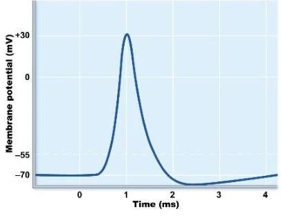
An action potential is a rapid reversal of the membrane potential caused by the opening of voltage-gated channels. The process follows a specific sequence:
Depolarization: The membrane becomes less negative (more positive) as cations enter the cell.
Repolarization: The membrane potential returns toward the resting level.
Hyperpolarization: The membrane potential briefly becomes more negative than the RMP.
Return to RMP: The cell stabilizes back to its resting state.
A graph illustrating the change in membrane potential over time, depicting an action potential.
Characteristics and Conduction
Action potentials are "all-or-none" events that occur once a specific threshold is reached. To communicate the intensity of a stimulus, the nervous system uses the frequency of action potentials rather than their size.
Conduction velocity (the speed of the signal) is influenced by:
Axon diameter: Larger diameters increase speed.
Myelination: Myelin allows for faster conduction via the Nodes of Ranvier.
During propagation, the neuron enters refractory periods. In the absolute refractory period, a second action potential is impossible. In the relative refractory period, a second action potential can occur, but only if the stimulus is stronger than usual (suprathreshold).
Chemical Synapses and Summation
When an action potential reaches an axon terminal, it triggers the release of neurotransmitters into the synaptic cleft. These chemicals bind to ligand-gated ion channels on the receiving cell, creating graded potentials. Unlike action potentials, graded potentials decay over time and distance.
EPSPs and IPSPs
The effect on the postsynaptic neuron depends on which channels open:
Excitatory Postsynaptic Potentials (EPSPs): Occur when cation channels open (mostly Na+ moving in), making the cell more likely to fire.
Inhibitory Postsynaptic Potentials (IPSPs): Occur when different channels open (mostly Cl− moving in), making the cell less likely to fire.
Summation
A single EPSP is often too weak to trigger an action potential. The neuron must use summation to reach threshold:
Temporal Summation: Multiple signals arriving at a single synapse in rapid succession.
Spatial Summation: Multiple signals arriving from different synapses at the same time.
Spinal Cord and Spinal Nerves
The spinal cord serves as the conduit between the brain and the body, housing complex pathways and reflex arcs.
Internal Structure
The spinal cord is organized into two distinct types of tissue:
White Matter: Located on the periphery, containing ascending (sensory) and descending (motor) tracts.
Sensory tracts include the dorsal column-medial lemniscal and the anterolateral/spinothalamic tracts.
Motor tracts include the pyramidal/corticospinal tracts.
Gray Matter: Located centrally, organized into dorsal horns (sensory) and ventral horns (motor).
Spinal Nerves and Plexuses
There are 31 pairs of spinal nerves. Each nerve is protected by connective tissue layers: the epineurium, perineurium, and endoneurium.
A typical spinal nerve is formed by the joining of a dorsal root (sensory) and a ventral root (motor). After exiting the spinal cord, the nerve divides into rami:
Dorsal ramus: Supplies the skin and deep muscles of the back.
Ventral ramus: Supplies the skin and muscles of the limbs and anterolateral chest.
Communicating rami: Connect the spinal nerve to the autonomic nervous system.
The ventral rami often merge to form complex networks called plexuses. Key plexuses include:
Cervical plexus: Gives rise to the phrenic nerve.
Brachial plexus: Supplies the arm (e.g., axillary, musculocutaneous, median, ulnar, and radial nerves).
Lumbar plexus: Supplies the lower abdomen and legs (e.g., obturator and femoral nerves).
Sacral plexus: Gives rise to the large sciatic nerve.
Clinical Correlations: Dermatomes and Reflexes
Dermatomes: These are specific areas of skin supplied by a single spinal nerve.
Spinal Cord Reflexes: These are rapid, involuntary responses to stimuli that occur at the level of the spinal cord, such as the stretch reflex and the tendon reflex.
Keep reading for free.
Create an account to finish this reading and make it part of your study routine.