Introduction to the Nervous System: Sensory and Motor Functions
This content provides a foundational overview of the nervous system, detailing its functional divisions into sensory and motor pathways, basic neuronal structure, and organizational principles of the Central and Peripheral Nervous Systems.
The nervous system is a complex, highly organized network designed to sense the environment, process information, and execute responses. To understand how we interact with the world—from feeling a cold breeze to pulling our hand away from a hot stove—we must look at the system through two lenses: its functional divisions (what it does) and its structural organization (how it is built).
Functional Divisions: Sensory vs. Motor
The nervous system is functionally split into two primary pathways: the sensory (afferent) division and the motor (efferent) division.
The Sensory (Afferent) Division
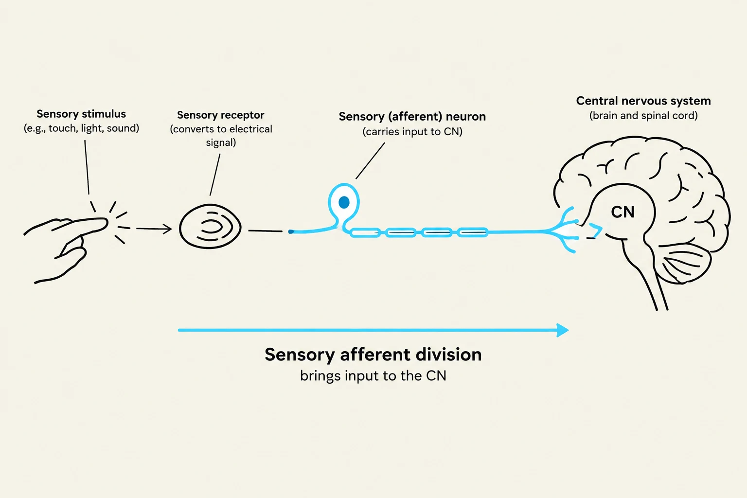
The sensory division is responsible for gathering input. It utilizes specialized receptors to detect stimuli (such as temperature, pressure, or light) and transmits this information toward the Central Nervous System (CNS). Think of this as the "input" side of the system.
The Motor (Efferent) Division
Once the CNS processes information, the motor division carries the "output" or action signals away from the CNS to effectors. Effectors are the structures that carry out the response, typically categorized as muscles (which move) or glands (which secrete).
Somatic and Visceral Subdivisions
Both the sensory and motor divisions are further categorized based on their target or origin:
Somatic System: Relates to the "soma" (body). It involves the skin, skeleton, and the voluntary control of skeletal muscles.
Visceral System: Relates to the internal organs (the "blood and guts"), such as the heart, lungs, and digestive tract.
Autonomic Nervous System: This is the involuntary motor component of the visceral system. It governs functions we cannot consciously control, such as cardiac muscle contraction, smooth muscle movement in tubular organs, and glandular secretions (like sweat or digestive enzymes).
Diagram illustrating the sensory afferent pathway, showing how sensory stimuli are detected by receptors, converted into electrical signals by sensory neurons, and transmitted to the central nervous system (brain and spinal cord).
Neuronal Structure and Classification
To facilitate these complex functions, the nervous system relies on specialized cells called neurons. A neuron's function is intimately tied to its structure, specifically its processes.
Basic Neuron Anatomy
Every neuron consists of three essential parts:
Dendrites: The receiving end that collects incoming information.
Cell Body (Soma): The central part that processes the incoming input and generates an output.
Axon: The long process that carries the output signal away to other neurons or effectors.
Classification by Structure and Function
Neurons are classified by the number of processes attached to the cell body (structure) and the direction in which they carry signals (function).
Functional Class
Structural Type
Description
Sensory Neurons
Typically Unipolar (or Bipolar)
Carry incoming signals from receptors to the CNS.
Motor Neurons
Always Multipolar
Carry outgoing signals from the CNS to effectors.
Interneurons
Always Multipolar
Located entirely within the CNS; they facilitate complex processing ("thinking").
Exam Tip: Remember that all interneurons and motor neurons are multipolar. If a question asks about a neuron located entirely within the CNS, it is an interneuron and must be multipolar.
System Organization: CNS and PNS
The nervous system is organized into two main structural regions: the Central Nervous System (CNS) and the Peripheral Nervous System (PNS).
Central Nervous System (CNS): Consists of the brain (the primary processing center) and the spinal cord (a relay station and reflex center).
Peripheral Nervous System (PNS): Consists of nerves and ganglia (clumps of cell bodies located outside the CNS).
Cranial Nerves: Emerge from the cranium to control the head, neck, and internal organs.
Spinal Nerves: Emerge from the spinal cord to control the body from the neck down.
The Reflex Arc: Speed Over Consciousness
A reflex is an unconscious, rapid response to a stimulus. Because reflexes can bypass the brain for immediate action, the body often responds to pain before the brain consciously perceives it.
In a typical reflex arc, a sensory neuron detects a stimulus (like a sharp thumbtack) and sends a signal to the spinal cord. An interneuron in the spinal cord may process this, or the sensory neuron may connect directly to a motor neuron. The motor neuron then triggers a muscle contraction to pull the limb away. This "shortcut" through the spinal cord is why you move your hand before you even realize you've been hurt.
Diagram illustrating the pathway of a sensory neuron, from stimulus detection by a sensory receptor to the transmission of a signal to the spinal cord via a unipolar sensory neuron and sensory ganglion.
Protection of the CNS
Because the CNS is vital to life and composed of delicate, non-mitotic cells (meaning they do not undergo mitosis to replace themselves), it requires extreme protection.
Physical Barriers
Bony Casings: The skull protects the brain, and the vertebral column protects the spinal cord.
Meninges: Three layers of connective tissue that anchor and protect the CNS.
Dura Mater: The "tough mother"; the outermost, durable layer.
Arachnoid Mater: The "spider mother"; a web-like middle layer.
Pia Mater: The "delicate mother"; the innermost layer that adheres closely to the brain and spinal cord.
Cerebrospinal Fluid (CSF): This fluid provides buoyancy (allowing the brain to "float"), cushions against impact, and helps clear metabolic waste products, especially during sleep.
The Blood-Brain Barrier (BBB)
To maintain a stable chemical environment, the brain utilizes a selectively permeable barrier formed by astrocytes (a type of glial cell) wrapping around capillaries. This ensures that only specific molecules can enter the brain tissue, protecting neurons from fluctuations in the blood.
Clinical Note: A concussion is essentially a bruising of the brain caused by the brain hitting the hard bony casing of the skull during impact. Repeated traumatic brain injuries can lead to long-term personality changes and structural damage.
Keep reading for free.
Create an account to finish this reading and make it part of your study routine.