An overview of the muscular system, covering its functions, types of muscle tissue (skeletal, cardiac, smooth), muscle organization, the neuromuscular junction, muscle attachments (origin, insertion, action), joint movements, and the connection between the muscular and circulatory systems.
The muscular system is a complex network of tissues responsible for much more than just lifting heavy objects. It is a fundamental component of human physiology that enables movement, maintains posture, stabilizes joints, and generates the heat necessary to maintain body temperature. Beyond these mechanical roles, different types of muscle tissue perform vital involuntary functions, such as the cardiac muscle pumping blood through the circulatory system or smooth muscle regulating the flow of substances through hollow organs.
Functions and Types of Muscle Tissue
The muscular system is categorized into three distinct types of tissue, each specialized for its specific location and role in the body:
Skeletal Muscle: These are long, cylindrical, striated fibers with many peripherally located nuclei. They are under voluntary control and are attached to bones to produce movement.
Cardiac Muscle: Found exclusively in the heart (myocardium), these cells are branched and striated. They function through involuntary, rhythmic contractions to pump blood.
Smooth Muscle: These spindle-shaped, non-striated cells lack visible striations and are controlled involuntarily. They are found in the walls of hollow organs, such as the gastrointestinal and respiratory tracts, where they regulate flow through processes like peristalsis or vasoconstriction.
Exam Tip: Remember that while skeletal and cardiac muscles are striated (showing a striped pattern under a microscope), smooth muscle is non-striated.
Muscle Organization and the Neuromuscular Junction
Skeletal muscle is organized in a hierarchical structure. A whole muscle is wrapped in a connective tissue sheath called the epimysium. Inside, the muscle is divided into bundles called fascicles (wrapped in perimysium), which are further composed of individual muscle fibers (cells) wrapped in endomysium. These fibers contain myofibrils, which are made of even smaller protein filaments called actin and myosin organized into units called sarcomeres.
Diagram illustrating the structure of a muscle, from the tendon to the myofibril, including its components like fascicles, muscle fibers, and sarcomeres with their respective bands and zones.
For a muscle to contract, it must receive a signal from the nervous system. This occurs at the neuromuscular junction (NMJ), a specialized synapse between a somatic motor neuron and a muscle fiber.
The process follows a specific sequence:
A nerve action potential reaches the axon terminal, causing voltage-gated Ca2+ channels to open.
Calcium influx triggers the release of the neurotransmitter acetylcholine (ACh).
ACh binds to receptors on the muscle's end plate, causing Na+ to enter the cell, which creates an end-plate potential (EPP).
This triggers a muscle action potential that travels into the fiber, leading to Ca2+ release from the sarcoplasmic reticulum and subsequent contraction.
A single motor neuron and all the muscle fibers it innervates constitute a motor unit. When the neuron fires, every fiber in that unit contracts simultaneously.
Origin, Insertion, and Muscle Action
To understand how muscles move the body, we must define their attachment points and their roles during movement:
Term
Definition
Origin
The more stable or proximal attachment; it remains fixed during contraction.
Insertion
The more mobile or distal attachment; it moves toward the origin during contraction.
Action
The specific joint movement produced by the contraction.
Muscles rarely work in isolation; they work in coordinated groups:
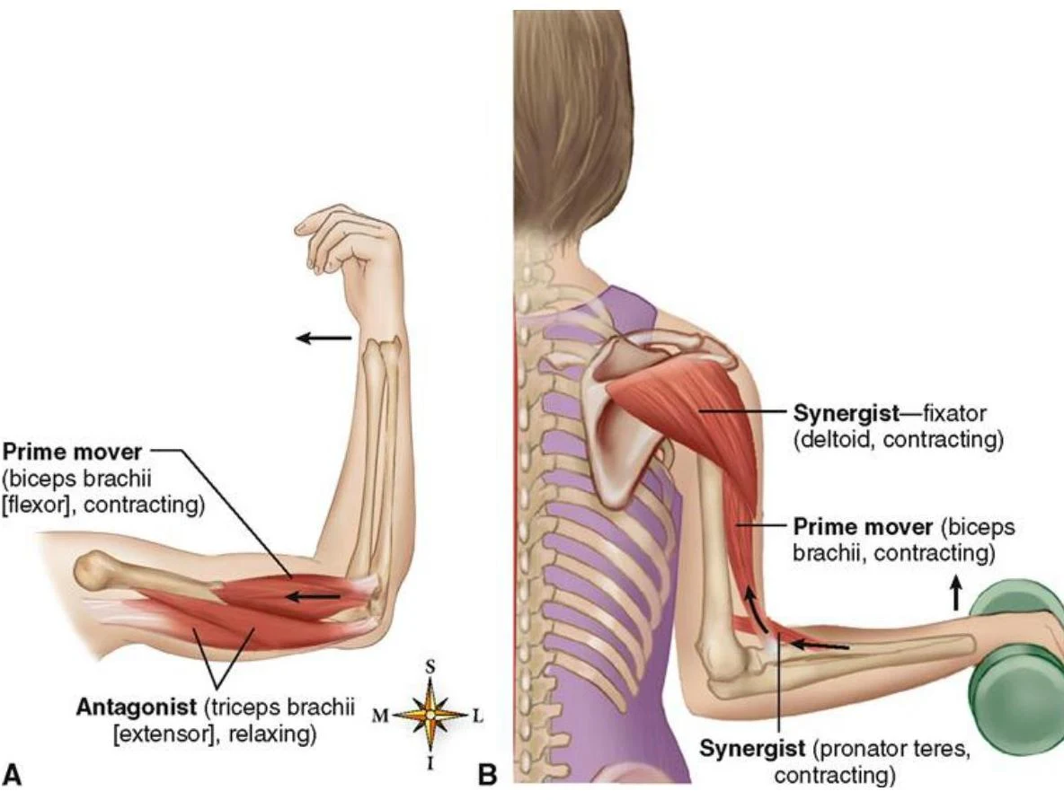
Agonist (Prime Mover): The main muscle responsible for a specific movement.
Antagonist: The muscle that opposes the agonist; it must lengthen or relax to allow the movement to occur.
Synergist: A muscle that assists the agonist by adding extra force or reducing unwanted motion.
Fixator: A muscle that stabilizes the origin of the agonist so the movement is efficient.
Diagrams illustrating the action of prime movers and synergist muscles during arm flexion and extension.
Synovial Joints and Movement
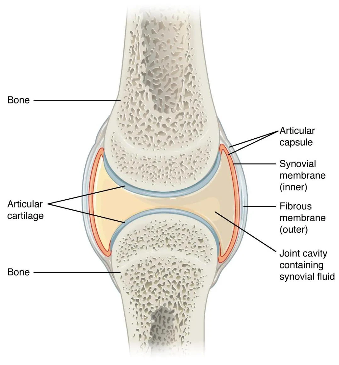
Muscles pull on bones, but the movement itself occurs at joints. Synovial joints are the most common and freely movable joints in the body. They are characterized by an articular capsule, articular (hyaline) cartilage to reduce friction, and a joint cavity filled with synovial fluid.
Diagram illustrating the anatomy of a synovial joint, including bone, articular cartilage, articular capsule, synovial membrane, fibrous membrane, and joint cavity containing synovial fluid.
Joint Types and Degrees of Freedom
Joints are classified by the number of planes in which they can move:
Biaxial joints: Allow movement in two planes (e.g., metacarpophalangeal joints of the fingers).
Multiaxial joints: Allow movement in three planes (e.g., the shoulder or hip).
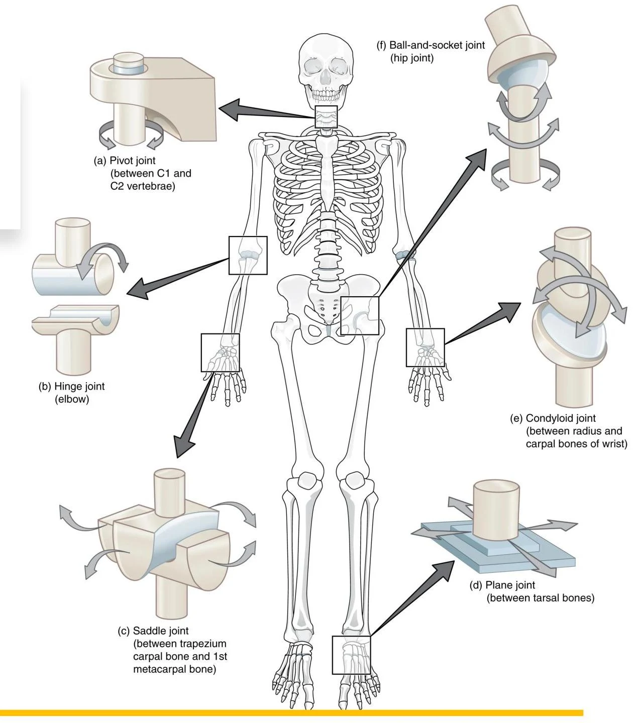
The six primary types of synovial joints include:
Pivot: Rotation (e.g., atlanto-axial joint).
Hinge: Flexion and extension (e.g., elbow, knee).
Saddle: Biaxial movement with opposition (e.g., the thumb).
Plane (Gliding): Small sliding movements (e.g., intercarpal joints).
Condylar (Ellipsoid): Flexion/extension and abduction/adduction (e.g., wrist).
Ball-and-Socket: Multiaxial movement (e.g., shoulder, hip).
This illustration shows different types of synovial joints in the human body, including pivot, hinge, saddle, ball-and-socket, condyloid, and plane joints, with examples of their locations.
Anatomical Directionality and Movement Terminology
To describe the body accurately, anatomists use specific directional terms, always assuming the subject is in the anatomical position.
Directional Terms
Superior/Inferior: Toward the head / Toward the feet.
Anterior/Posterior: Toward the front / Toward the back.
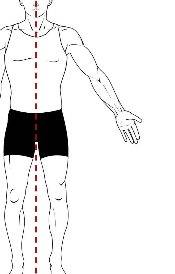
Medial/Lateral: Toward the midline / Away from the midline.
Proximal/Distal: Nearer to the trunk / Farther from the trunk.
Superficial/Deep: Closer to the surface / Farther from the surface.
Common Body Movements
Understanding movement requires knowing how joint angles change:
Flexion vs. Extension: Flexion decreases the angle between segments (e.g., bending the elbow), while extension increases it.
Abduction vs. Adduction: Abduction moves a limb away from the midline, while adduction moves it toward the midline.
Rotation: Medial rotation turns a surface toward the midline; lateral rotation turns it away.
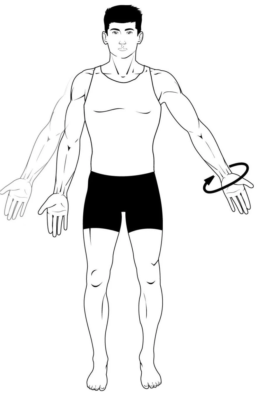
Circumduction: A circular movement of a limb while the rest of the body stays still.
Specialized Movements: These include dorsiflexion/plantar flexion (foot), inversion/eversion (foot), and supination/pronation (forearm).
An illustration of the human body with a dashed line indicating the midline.
Illustration showing wrist rotation.
The Muscular-Circulatory Connection
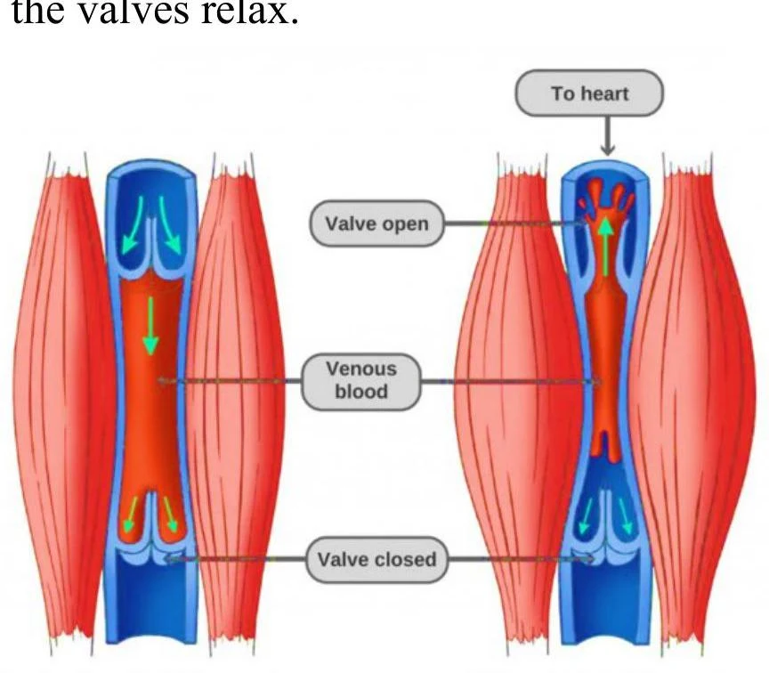
The muscular system is deeply connected to the circulatory system. While the heart (cardiac muscle) acts as the primary pump, skeletal muscles play a crucial role in venous return. Because veins in the lower body must fight gravity, they rely on the "skeletal muscle pump." When skeletal muscles contract, they compress the veins, pushing blood through one-way valves toward the heart.
Diagram illustrating the function of venous valves in the leg muscles, showing how they open to allow blood flow towards the heart and close to prevent backflow.
Keep reading for free.
Create an account to finish this reading and make it part of your study routine.