An overview of the endocrine system, its chemical messengers (hormones), and their mechanisms of action. Explains the roles of the hypothalamus and pituitary gland in regulating bodily functions through hormone secretion and feedback systems.
The human body relies on a complex communication network to maintain internal stability, a state known as homeostasis. While the nervous system provides rapid electrical signaling, the endocrine system provides a slower, more sustained chemical communication method by secreting messengers called hormones directly into the bloodstream. Together, these two systems form the neuroendocrine system, which integrates the body's response to both internal changes and the external environment.
Chemical Messengers: Hormones and Receptors
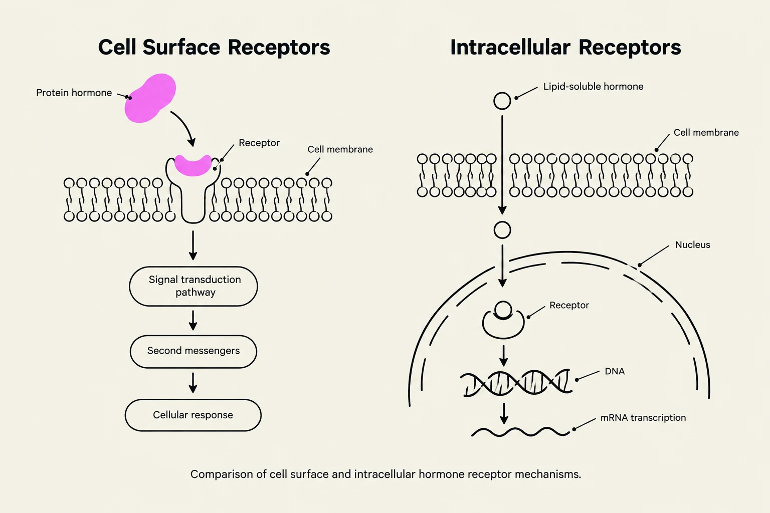
Comparison of cell surface and intracellular hormone receptor mechanisms.
Hormones are chemical messengers produced by glands—organized groups of specialized cells that lack ducts, meaning they must secrete their products directly into the blood. Because they travel through bodily fluids, hormones are present in the body at all times, though their concentrations fluctuate based on physiological needs.
Classification by Structure
Hormones are classified into three primary chemical categories:
Proteins and Polypeptides: The most prominent type.
Steroids: Lipid-based hormones.
Amines: Derived from amino acids.
Mechanism of Action
To exert an effect, a hormone must bind to a specific receptor site on or within a target cell. The location of these receptors determines how the hormone functions:
Cell Surface Receptors: These hormones bind to receptors on the exterior of the cell membrane, triggering a response via G proteins or enzyme systems.
Intracellular Receptors: Lipid-soluble (nonpolar) hormones can pass through the cell membrane to bind with receptors inside the cytoplasm or the nucleus. Once the hormone-receptor complex forms, it can bind to specific sequences of DNA to activate or repress the transcription of genes into messenger RNA (mRNA).
The Master Controller: The Hypothalamus
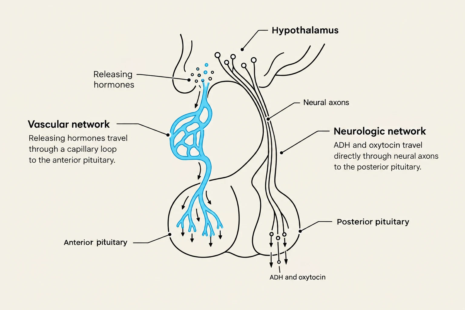
Diagram illustrating the relationship between the hypothalamus and the pituitary gland, detailing the vascular and neurologic networks involved in hormone transport.
The hypothalamus acts as the coordinating center for the neuroendocrine system. It functions as a "master gland" by constantly monitoring homeostasis through the analysis of input from the central nervous system (CNS) and the periphery. It regulates vital functions such as body temperature and thirst.
The hypothalamus communicates with the pituitary gland through the hypothalamic-pituitary axis (HPA) using two distinct pathways:
Vascular Network: A capillary network carries releasing hormones (such as TRH, GnRH, and CRH) or inhibiting factors (such as somatostatin) directly to the anterior pituitary.
Neurologic Network: A neural connection delivers antidiuretic hormone (ADH) and oxytocin to the posterior pituitary, where they are stored until needed.
The Pituitary Gland
Located within the bony sella turcica of the skull, the pituitary gland is divided into three distinct lobes, each with unique functions.
The Anterior Pituitary
The anterior lobe produces six major hormones that are essential for growth, reproduction, and metabolism. These hormones are often released in a rhythmic manner, following a diurnal rhythm (varying with sleep/wake cycles and light exposure) or in response to exercise.
A helpful mnemonic to remember these is FLAT PEG:
Follicle-stimulating hormone (FSH): Stimulates estrogen and progesterone in females, and testosterone in males.
Luteinizing hormone (LH): Also regulates estrogen, progesterone, and testosterone.
Adrenocorticotropic hormone (ACTH): Triggers the release of glucocorticoids and androgens; it is vital for the body's acute stress response.
Prolactin (PRL): Stimulates milk production and secretion.
Endorphins: Produced in the intermediate lobe.
Growth hormone (GH): Promotes cell growth.
The Posterior Pituitary
Unlike the anterior lobe, the posterior pituitary does not produce its own hormones; instead, it stores and releases two hormones produced by the hypothalamus:
Antidiuretic Hormone (ADH/Vasopressin): Released when blood osmolarity increases or blood volume decreases. It acts on the kidneys to increase water retention.
Oxytocin: Regulates uterine contractions during late pregnancy and the "let-down" reflex for milk release during lactation.
The Intermediate Lobe
The intermediate lobe produces endorphins and enkephalins, which are released in response to severe pain or stress to modulate pain receptors. It also secretes Melanocyte-stimulating hormone (MSH).
Exam Tip: The "Fight-or-Flight" Response
If you are asked which hormone prepares the body for an acute stress response, the answer is ACTH. Activation of the sympathetic nervous system directly triggers the release of ACTH to prepare the body for action.
Endocrine Regulation and Feedback Systems
To maintain precise hormone levels, the body utilizes various regulatory mechanisms.
Negative Feedback Systems
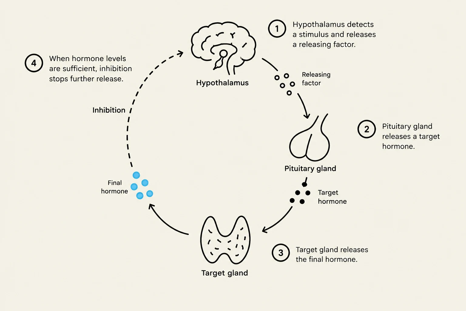
This diagram illustrates the hormonal regulation of a target gland, starting with the hypothalamus releasing a factor that stimulates the pituitary gland. The pituitary then releases a target hormone, which acts on the target gland to produce a final hormone. High levels of the final hormone trigger inhibition, stopping further release.
Most endocrine activity is fine-tuned through negative feedback systems, which function similarly to the law of supply and demand:
The hypothalamus senses a need for a hormone and secretes a releasing factor.
The pituitary responds by secreting the target hormone.
The target gland produces the final hormone.
Once the hypothalamus senses that hormone levels have risen sufficiently, it stops secreting the releasing factor to prevent overproduction.
Direct Stimuli
Not all hormones are controlled by the hypothalamus. Some are released in response to direct physiological changes in the environment:
Parathyroid Hormone: Released by the parathyroid glands in response to local calcium levels.
Renin and Erythropoietin: Released by the kidneys in response to changes in blood flow and oxygenation.
Insulin, Glucagon, and Somatostatin: Produced by the pancreas in response to varying blood glucose levels.
References
CH 34 Introduction to the Endocrine System.pptx (Source 1)
Karch’s Focus on Nursing Pharmacology, 10th ed. (Reference)
Keep reading for free.
Create an account to finish this reading and make it part of your study routine.