Explains the structure and function of the digestive system, including the alimentary canal, accessory organs, and the processes of mechanical and chemical digestion.
The digestive system is a complex biological network designed to perform a single, vital task: the breakdown of food into forms that body cells can absorb. This process is not merely about crushing food; it involves two distinct but coordinated mechanisms: mechanical digestion, which physically breaks food into smaller pieces, and chemical digestion, which uses enzymes and acids to break down molecular bonds. To achieve this, the system utilizes a continuous pathway known as the alimentary canal and several accessory organs that provide essential secretions.
The Structure of the Alimentary Canal
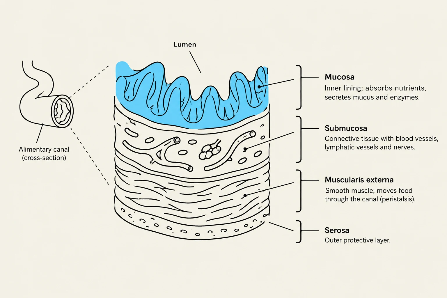
The alimentary canal is a continuous tube extending from the mouth to the anus. While the organs within this tube vary in function, they all share a similar structural blueprint consisting of four distinct layers:
Mucosa: The innermost layer, composed mostly of epithelial tissue. It is responsible for secreting mucus into the lumen (the passageway) and is highly active in absorbing nutrients.
Submucosa: Located just outside the mucosa, this layer contains loose connective tissue, blood vessels, lymphatic vessels, glands, and nerves. The vessels here are critical for transporting absorbed nutrients throughout the body.
Muscularis externa: A layer of smooth muscle tissue that facilitates movement through contractions, such as peristalsis, to push material through the tract.
Serosa: The outermost protective layer. In certain areas, such as the abdominal cavity, this is known as the peritoneum.
Diagram illustrating the layers of the alimentary canal: mucosa, submucosa, muscularis externa, and serosa.
Exam Tip: Remember that the submucosa is the "transportation hub" because it houses the blood and lymphatic vessels that carry nutrients away from the site of absorption.
The Alimentary Pathway: From Mouth to Stomach
The Mouth and Pharynx
Digestion begins in the mouth through mastication (chewing). Teeth—including incisors, canines, premolars, and molars—work together to reduce food size. Simultaneously, chemical digestion begins as the salivary glands secrete saliva containing salivary amylase, an enzyme that initiates the breakdown of carbohydrates. The tongue mixes this food with saliva to form a soft mass called a bolus.
Once swallowed, the bolus passes through the pharynx. Swallowing is largely a reflex action where the soft palate moves to prevent food from entering the nasal cavity, and the epiglottis covers the larynx to prevent food from entering the airway. The bolus then enters the esophagus, a muscular tube that uses peristalsis to move food toward the stomach. Entry into the stomach is regulated by the lower esophageal sphincter (also called the cardiac sphincter), which acts like a valve to prevent stomach contents from refluxing into the esophagus.
The Stomach
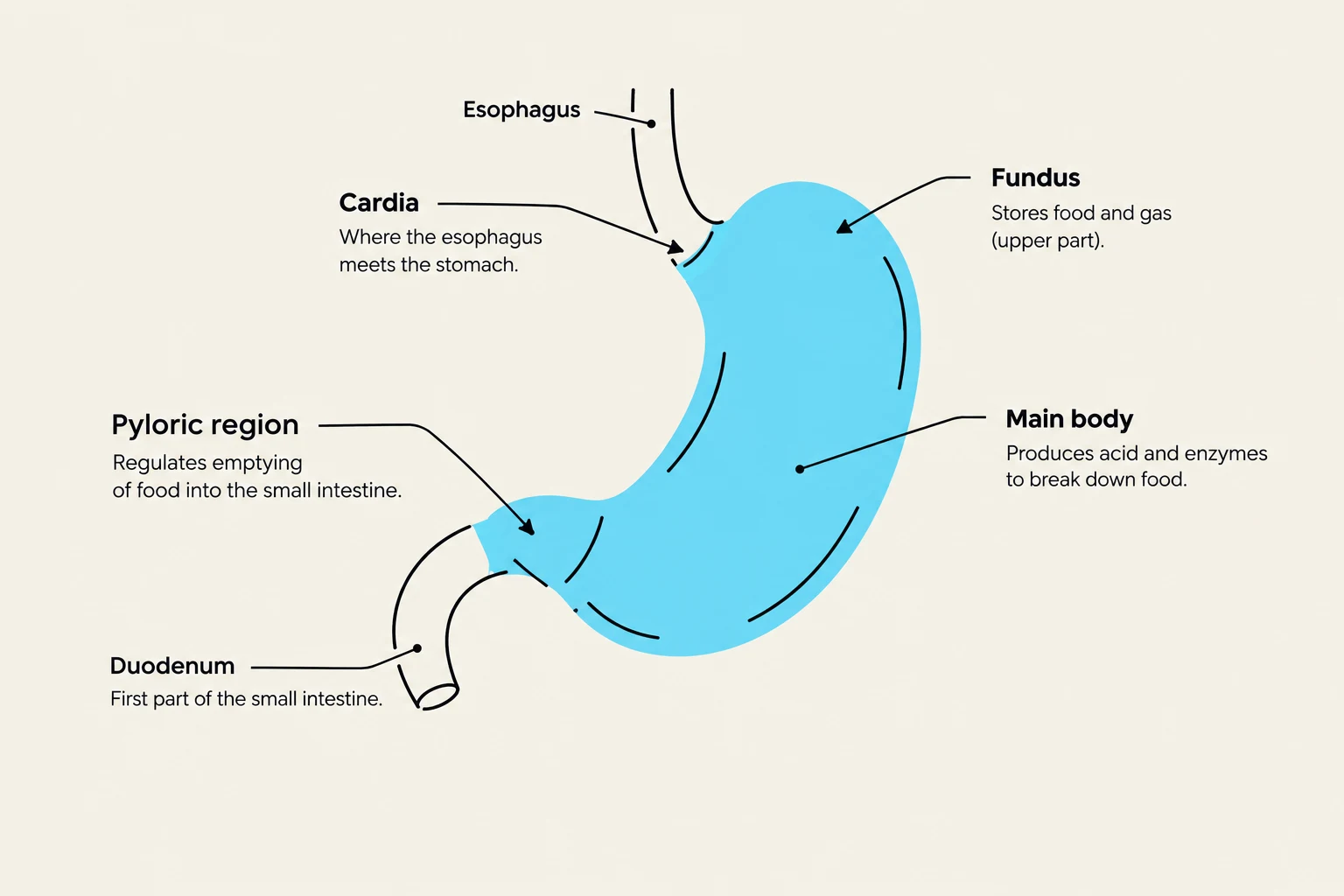
The stomach is a sac-like organ located in the upper left quadrant of the abdomen. Here, food is mixed with gastric juice to create a semi-liquid mixture called chyme. The stomach's lining contains gastric glands with specialized cells:
Mucous cells: Secrete mucus to protect the lining.
Gastric cells (Chief cells): Secrete pepsinogen, which is converted into the active enzyme pepsin by hydrochloric acid to begin protein digestion.
Parietal cells: Secrete hydrochloric acid (HCl) and intrinsic factor, the latter of which is essential for the absorption of vitamin B12.
Diagram of the human stomach and its parts: Esophagus, Cardia, Fundus, Main body, Pyloric region, and Duodenum.
Nutrient Absorption: The Small and Large Intestines
The Small Intestine
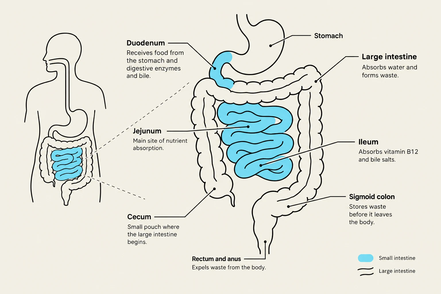
The small intestine is the primary site of nutrient absorption. It is divided into three sections:
Duodenum: The first, relatively large portion where chyme meets digestive enzymes.
Jejunum: The middle, highly coiled section.
Ileum: The final section that attaches to the large intestine.
To maximize efficiency, the small intestine utilizes villi and microvilli to vastly increase its surface area for absorption. Various enzymes, such as maltase, lactase, and peptidases, complete the breakdown of sugars and proteins.
The Large Intestine
The large intestine receives the remaining material from the ileum at the cecum. It consists of the ascending colon, transverse colon, descending colon, and sigmoid colon. The primary role of the large intestine is to absorb water and electrolytes from the remaining material, turning it into solid feces. Feces are stored in the rectum before being eliminated through the anal canal and anus.
Diagram illustrating the human digestive system, including the stomach, small intestine (duodenum, jejunum, ileum), and large intestine (cecum, sigmoid colon, rectum, and anus).
The Role of Accessory Organs
While food does not pass through them, accessory organs are indispensable for chemical digestion:
Salivary Glands: Produce saliva to moisten food and start carbohydrate breakdown.
Liver: A large organ that produces bile, a substance essential for the emulsification (breaking down) of fats.
Gallbladder: A small sac that stores and concentrates bile before releasing it into the duodenum.
Pancreas: Secretes "pancreatic juice" containing enzymes like lipases (for fats), proteases (for proteins), and amylases (for carbohydrates), as well as bicarbonate to neutralize the acidic chyme coming from the stomach.
Summary of Absorbed Nutrients
The ultimate goal of this entire process is the absorption of essential nutrients into the bloodstream:
Carbohydrates: Broken down into simple sugars like glucose.
Lipids: Broken down into triglycerides and cholesterol.
Proteins: Broken down into amino acids.
Vitamins and Minerals: Essential micronutrients (e.g., Calcium, Iron, Vitamin B12).
Water: Absorbed primarily in the large intestine.
Keep reading for free.
Create an account to finish this reading and make it part of your study routine.