In the complex world of biology, proteins serve as the essential "workforce" of the cell. They are responsible for a vast array of tasks, from providing structural support to acting as enzymes that catalyze chemical reactions. The most critical rule to remember when studying proteins is that structure determines function. This means that the specific three-dimensional shape a protein assumes is what allows it to perform its unique biological role. If a protein loses its shape, it often loses its ability to function.
The Building Blocks: Amino Acids
Proteins are polymers, which means they are large molecules made of repeating smaller units called monomers. For proteins, these monomers are 20 different types of amino acids.
Amino Acid Structure
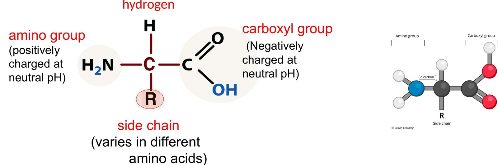
Every amino acid shares a common chemical framework centered around a central carbon atom. Attached to this central carbon are four distinct components:
An amino group (−NH2)
A carboxyl group ($-COOH$), which is negatively charged at neutral pH
A hydrogen atom
A variable side chain, also known as the R group
Diagram illustrating the general structure of an amino acid, showing the amino group, carboxyl group, hydrogen atom, and a variable side chain (R group).
The R group is the most important part of the amino acid for protein diversity because it is unique to each of the 20 types. The chemical properties of these side chains dictate how the amino acids will interact with one another.
Categorizing Side Chains
Based on the chemical nature of the R group, amino acids can be classified into three main categories:
Nonpolar side chains: These typically consist of carbon (C), hydrogen (H), and sometimes sulfur (S). They are hydrophobic, meaning they tend to avoid water.
Polar uncharged side chains: These may have partial positive (δ+) or partial negative (δ−) charges.
Polar charged side chains: These possess a full positive ($+$) or negative ($-$) charge.
Study Tip: To quickly identify a side chain's property, look for oxygen (O) or nitrogen (N) atoms, which often indicate polarity, or look for full charges. If the side chain contains only C, H, and S, it is almost certainly nonpolar.
Linking Amino Acids: Polymerization
To build a protein, amino acids must be linked together into a chain. This process is called polymerization.
Condensation and Hydrolysis
The bonds between amino acids are formed through condensation reactions (also known as dehydration reactions). During this process, a water molecule (H2O) is removed to join the monomers together. Specifically, the carboxyl group of one amino acid joins with the amino group of the next, creating a covalent bond known as a peptide bond.
This illustration shows the dehydration synthesis reaction between two amino acids to form a peptide bond and a molecule of water.
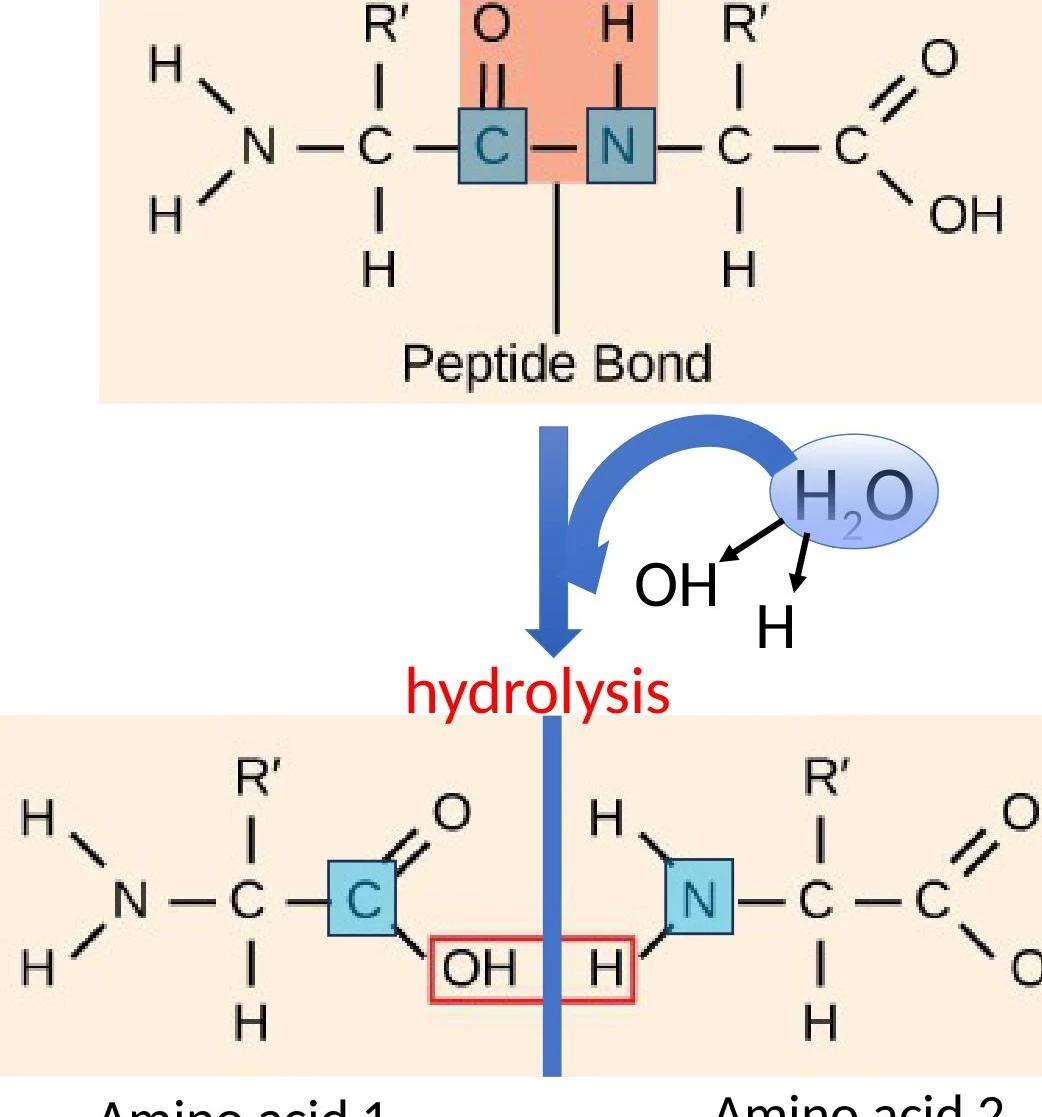
Conversely, when the cell needs to break these bonds, it performs a hydrolysis reaction. In hydrolysis, a water molecule is added back into the structure, which breaks the peptide bond by adding an $-OH$ group to the carbon of the first amino acid and an −H to the nitrogen of the second.
This diagram illustrates the process of hydrolysis, where a peptide bond is broken by the addition of a water molecule, resulting in the formation of two amino acids.
The Four Levels of Protein Structure
A long chain of amino acids, called a polypeptide, does not stay as a simple string. It must fold into a complex shape to become a functional protein. This folding is organized into four hierarchical levels:
1. Primary Structure
The primary structure is the linear sequence of amino acids in the polypeptide chain. This sequence is held together by the peptide bonds mentioned earlier. The specific order of amino acids is vital; if even one amino acid is changed due to a mutation, the entire protein might fail to fold correctly, leading to a loss of function.
2. Secondary Structure
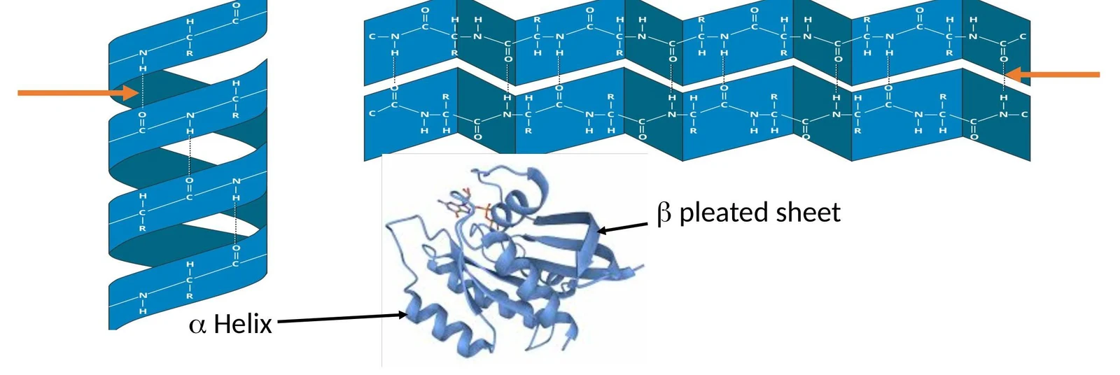
As the chain grows, local regions begin to fold into specific patterns due to hydrogen bonds forming between the atoms of the polypeptide backbone. The two most common types of secondary structure are:
Diagrams illustrating the alpha helix and beta pleated sheet structures of proteins.
3. Tertiary Structure
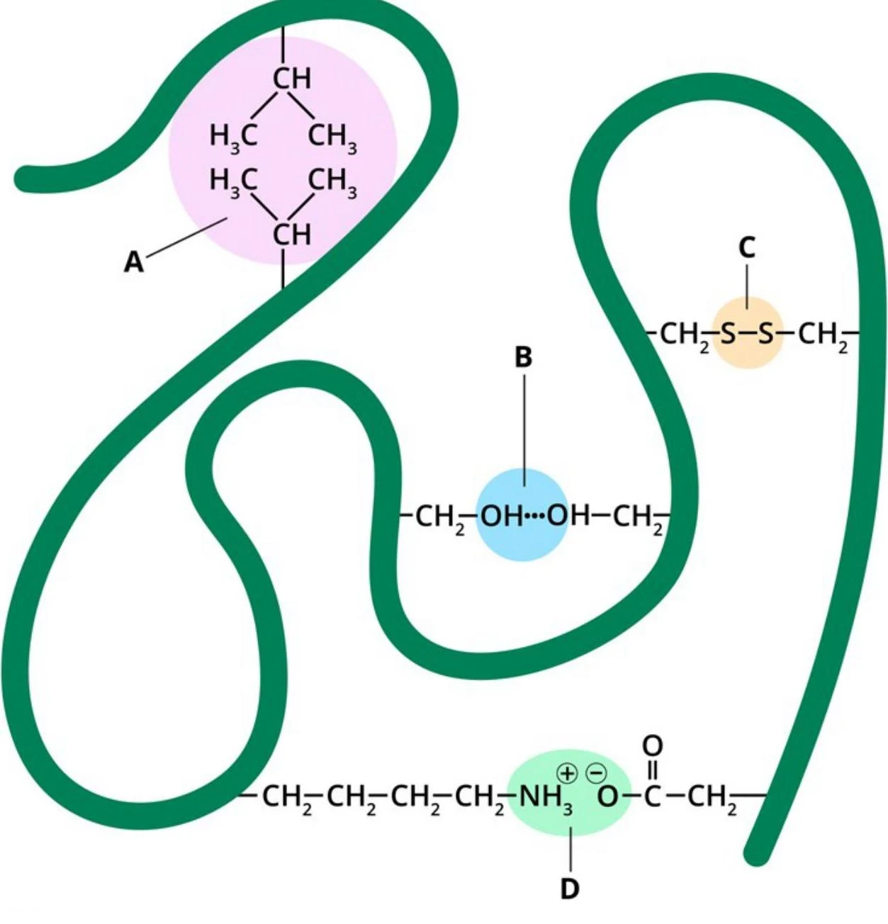
The tertiary structure is the overall three-dimensional conformation of a single polypeptide chain. This level of structure is determined by various interactions between the R groups (side chains) of the amino acids. These interactions include:
Hydrophobic interactions (nonpolar side chains clustering together to avoid water)
Hydrogen bonds
Ionic bonds
Disulfide bridges
This illustration depicts various functional groups and bonds found in amino acids and proteins, including hydrophobic side chains (A), hydrogen bonding (B), disulfide bonds (C), and charged amino and carboxyl groups (D).
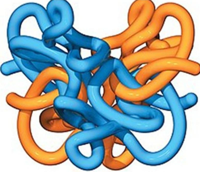
4. Quaternary Structure
Some functional proteins consist of more than one polypeptide chain working together. When multiple subunits aggregate into a single functional complex, it is referred to as the quaternary structure.
A representation of a protein structure, possibly hemoglobin, showing intertwined polypeptide chains in blue and orange.
Protein Folding and Chaperones
Protein folding is the process by which a polypeptide achieves its functional shape. Because the environment inside a cell is crowded, proteins sometimes struggle to fold correctly on their own.
To assist in this, cells utilize specialized molecules called chaperone proteins. Chaperones guide partially folded proteins to help them achieve their correct three-dimensional conformation and prevent them from forming improper, non-functional shapes.
Clinical Connection: When proteins misfold, they can cause serious health issues. For example, insulin is a protein hormone that regulates blood sugar. If a mutation in the primary structure causes insulin to misfold, it cannot signal cells to take up glucose, which can lead to unregulated blood sugar levels and diabetes.
Keep reading for free.
Create an account to finish this reading and make it part of your study routine.