Introduction to Musculoskeletal Kinesiology and Biomechanics
Explore the foundational concepts of kinesiology and biomechanics, bridging the gap between anatomical structure and functional occupation. Understand how the human body moves through an integrated approach of physical, cognitive, and psychological factors.
Understanding how the human body moves requires a multi-dimensional approach. It is not enough to simply know the names of muscles or bones; one must understand how these structures interact with physics and psychology to allow a person to engage in their daily lives. This article explores the foundational concepts of kinesiology and biomechanics, bridging the gap between anatomical structure and functional occupation.
The Holistic View of Function
To understand movement in a clinical setting, we often utilize the ICF Framework (International Classification of Functioning, Disability and Health). This framework views functioning through three primary lenses: body structures and functions, activities, and participation.
Furthermore, human performance exists at a Physical-Cognitive-Psychological Intersection. A person's ability to perform an occupation is rarely dictated by physical strength alone. It is a combination of:
Physical: Strength, endurance, and posture.
Cognitive: Attention, processing speed, and problem-solving.
Psychological: Motivation and emotional regulation.
A limitation in one of these areas—such as a lack of motivation (psychological) or poor attention (cognitive)—can significantly impact a person's ability to utilize their physical strength to complete a task.
Kinesiology: The Language of Motion
Kinesiology involves the study of movement, which can be broken down into two distinct categories: Osteokinematics and Arthrokinematics.
Osteokinematics: This refers to the visible movement of bones in space (e.g., swinging your arm up during shoulder flexion).
Arthrokinematics: This refers to the microscopic movements occurring at the joint surfaces themselves, such as gliding or rolling.
Planes and Axes of Motion
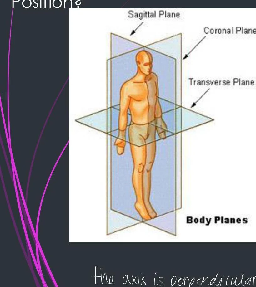
To describe osteokinematics accurately, we use specific anatomical planes and axes. The axis of motion is always perpendicular to the plane of motion.
Sagittal Plane: Splits the body into right and left sides. Movements include flexion and extension (e.g., elbow flexion).
Frontal (Coronal) Plane: Splits the body into anterior (front) and posterior (back) parts. Movements include abduction (moving away from midline) and adduction (moving toward midline).
Transverse (Axial) Plane: Splits the body into upper and lower parts. Movements include rotation and horizontal abduction/adduction.
An illustration showing the sagittal, coronal, and transverse planes of the human body.
Directional Terminology
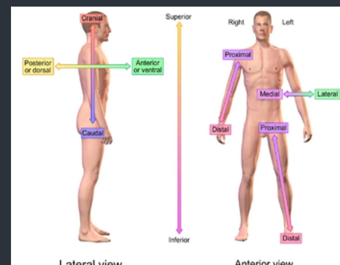
Navigating the body requires a standardized vocabulary to describe where structures are located relative to one another:
Term
Definition
Term
Definition
Cranial
Towards the head
Medial
Towards the midline
Caudal
Towards the "tail"/bottom
Lateral
Away from the midline
Superior
Above
Anterior/Volar
Towards the front
Inferior
Below
Posterior/Dorsal
Towards the back
Proximal
Towards the body center
Distal
Away from the body center
Anatomical directional terms are illustrated on anterior and lateral views of the human body, indicating cranial, caudal, superior, inferior, anterior, posterior, medial, lateral, proximal, and distal positions.
Muscle Contractions and Kinetic Chains
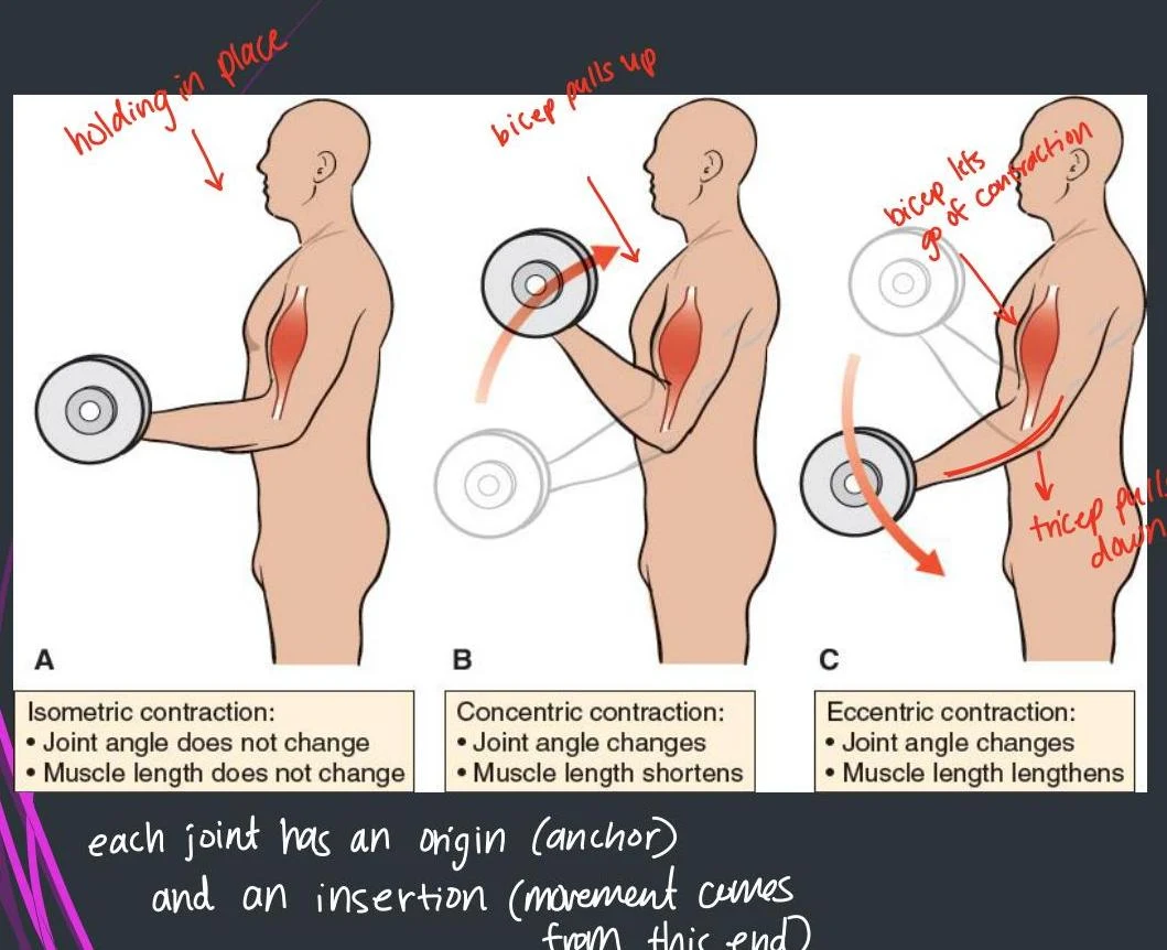
When muscles produce force, they do so in different ways depending on whether the muscle length or the joint angle is changing.
Isometric Contraction: The muscle produces force, but the joint angle and muscle length remain unchanged (e.g., holding a heavy grocery bag steady at your side).
Concentric Contraction: The muscle shortens as it overcomes resistance, changing the joint angle (e.g., the upward phase of a bicep curl).
Eccentric Contraction: The muscle lengthens while under tension, controlling the movement (e.g., slowly lowering a weight).
Diagram illustrating isometric, concentric, and eccentric muscle contractions during a bicep curl, with accompanying text explaining joint origin and insertion points.
Open vs. Closed Kinetic Chains
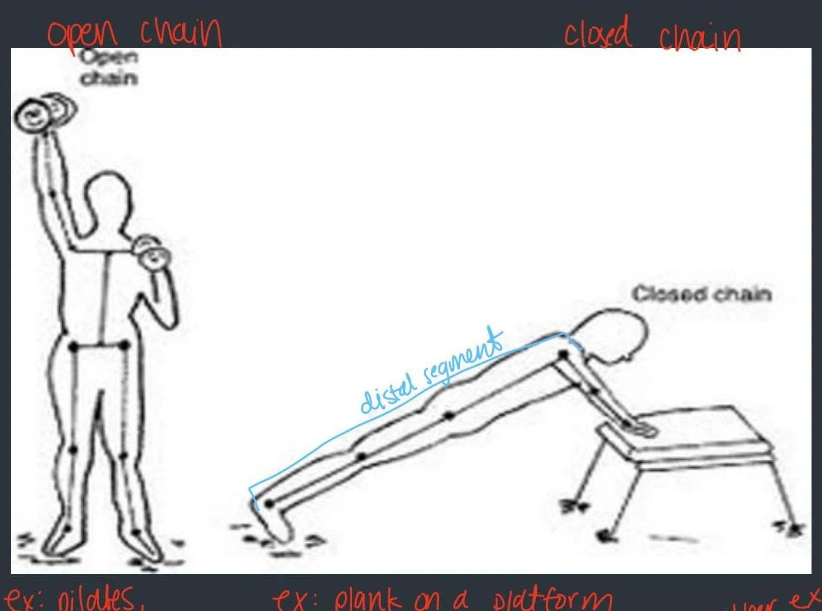
We also categorize movements based on whether the end of the limb is free or fixed:
Open Chain: The distal segment (the part furthest from the body, like the hand) is free to move. Example: Self-feeding with a spoon.
Closed Chain: The distal segment is fixed against a surface. Example: A sit-to-stand transfer where the feet are planted on the floor.
Diagram illustrating open chain and closed chain exercises, with examples of Pilates and planks.
Biomechanics: Forces and Levers
Biomechanics applies the laws of physics to human movement. Two primary forces we encounter are tensile force (pulling) and compressive force (pushing). For example, the vertebral bodies are crucial for absorbing compressive forces when lifting heavy objects.
The human body operates using three types of levers, which are simple machines designed to either overcome resistance, provide balance, or increase speed/distance.
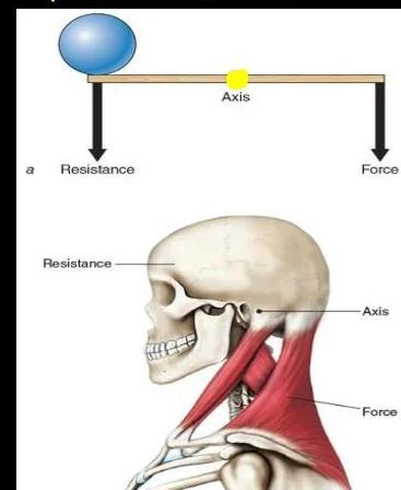
First-Class Lever: The axis (fulcrum) is between the force and the resistance. This system is primarily used for balance, such as the movement of the head on the neck.
Diagrams illustrating the principles of levers and forces in the human body, specifically the neck muscles acting as a force to move the head against resistance.
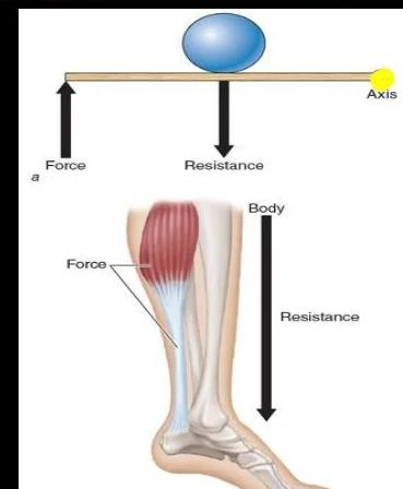
Second-Class Lever: The resistance is in the middle, with the axis at one end and the force at the other. This allows for overcoming large resistance with less effort, such as the calf muscle during a toe raise.
Diagrams illustrating the principles of force, resistance, and axis in mechanical systems, including a lever and a human leg muscle.
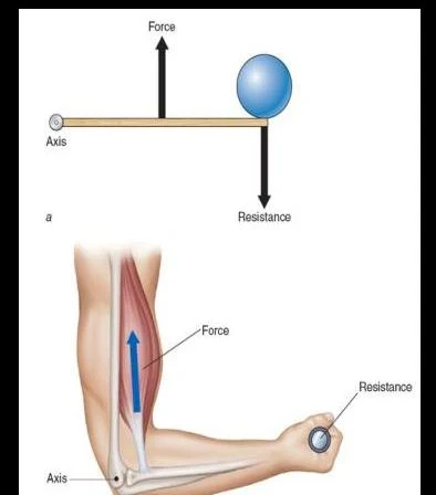
Third-Class Lever: The force is applied in the middle, between the axis and the resistance. This is the most common lever in the human body (e.g., the elbow joint) and is designed to increase speed and distance.
Diagrams illustrating the principles of levers and forces in the human arm.
Exam Tip: When identifying a lever, always locate the Axis (the pivot point), the Force (the muscle contraction), and the Resistance (the weight or gravity). The order of these three components determines the class of the lever.
Neurological Foundations
Movement is not just mechanical; it is driven by the nervous system. The Cranial Nerves (CN I-XII) are essential for sensory and motor functions in the head and neck. For instance, the Hypoglossal nerve (CN XII) is responsible for moving the tongue, which is critical for speech and swallowing.
To assess neurological health, clinicians use:
Dermatomes: Areas of skin supplied by a single spinal nerve root (sensory).
Myotomes: Groups of muscles supplied by a single spinal nerve root (motor).
Understanding these maps allows clinicians to localize exactly where a neurological injury may have occurred based on where a patient feels sensation or lacks strength.
Keep reading for free.
Create an account to finish this reading and make it part of your study routine.