Introduction to Microscopes and Biological Measurements
Explore the fundamental principles of microscopy, including the types of microscopes, their components, and optical properties like magnification and resolution. Learn how to perform biological measurements using microscopy techniques.
In the study of life, the microscope is perhaps the most transformative instrument ever developed. Most biological structures, particularly cells, fall below the limit of resolution of the human eye, which is approximately 0.1 mm. To observe these microscopic worlds, scientists rely on various types of microscopes that use different sources of illumination to reveal detail and structure.
Types of Microscopes
Microscopes are primarily classified by their source of illumination.
Light Microscopes
Light microscopes use visible light and a sequence of lenses to magnify specimens.
Dissecting Microscope: Designed for viewing larger, three-dimensional specimens. It features a stage and switches that allow for both above-specimen and below-specimen illumination.
Compound Light Microscope: This is a more powerful tool that uses light passing through a specimen and a series of lenses to form a magnified image. It can achieve magnifications up to 2,500× and has a resolution of 0.2μm (without the use of immersion oil).
Electron Microscopes
While light microscopes are limited by the wavelength of visible light, electron microscopes use electron beams with much shorter wavelengths. This allows them to achieve much higher resolution, enabling the visualization of sub-cellular structures as small as 0.2 nm.
Anatomy of a Compound Microscope
To use a microscope proficiently, one must understand its mechanical and optical components.
Magnification Components
Ocular Lens: The eyepiece lens that the user looks through to see the magnified image.
Objective Lenses: A series of lenses mounted on a rotating nosepiece located just above the stage. These provide different levels of magnification (e.g., 4×, 10×, 40×).
Illumination and Focus
Condenser: A lens system that focuses light into a beam of parallel waves onto the specimen.
Iris Diaphragm: A mechanism that regulates the amount of light passing through the condenser. Reducing the light can actually increase the visibility of semi-transparent objects by improving contrast.
Mechanical Stage: The flat surface that supports the slide. In compound microscopes, this stage often includes mechanical stage knobs to move the slide precisely.
Focus Knobs: The coarse focus knob is used for initial, rough adjustments to the distance between the lens and the specimen. The fine focus knob is used for precise, sharp adjustments, especially at higher magnifications.
Exam Tip: Total Magnification
To find the total magnification of your view, always multiply the magnification of the ocular lens by the magnification of the objective lens currently in use.
Total Magnification=Ocular Magnification×Objective Magnification
Optical Principles: Resolution, Magnification, and Depth of Field
Understanding the physics of light is essential for interpreting what you see through the lens.
Resolution
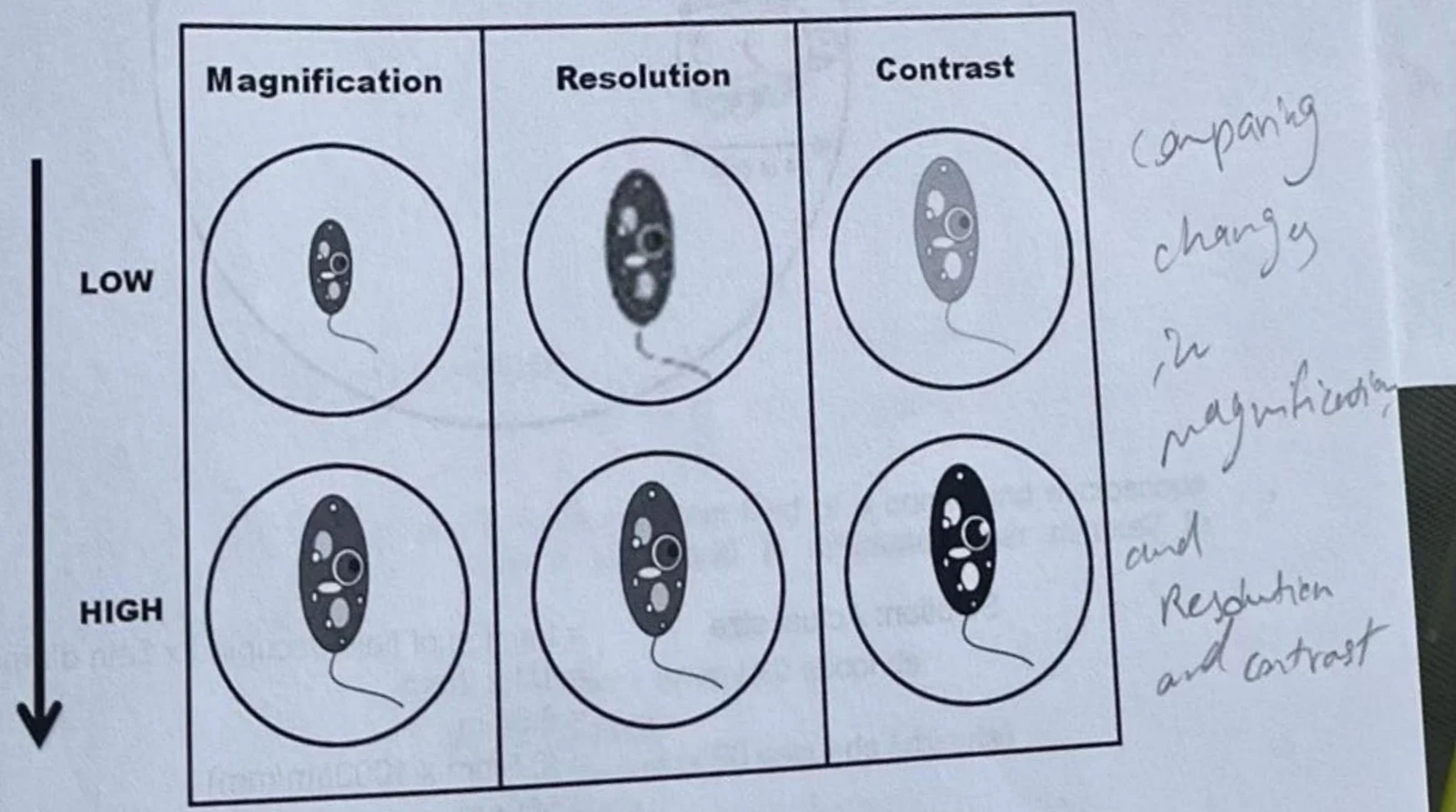
Resolution is the ability to distinguish between two points as separate entities. A high-resolution image is sharp, whereas a low-resolution image is fuzzy, regardless of how much it is magnified. Resolution can be improved by:
Using a blue filter: Shorter wavelengths of light (like blue) reduce diffraction and increase resolution.
Immersion Oil: Using a special oil with a 100× objective lens decreases light diffraction, allowing more light to enter the lens.
Depth of Field
The depth of field refers to the vertical thickness of the specimen that remains in focus at a single time. There is an inverse relationship between magnification and depth of field: as magnification increases, the depth of field decreases.
Diagram illustrating the effects of magnification, resolution, and contrast on the appearance of a microscopic organism.Figure 1-3. Comparison of the effect of magnification, resolution, and contrast on a microscopic image.
Biological Measurements
Microscopy allows for the quantification of biological specimens through several mathematical approaches.
1. Estimating Specimen Size
You can calculate the actual size of a specimen by comparing how much of the "field of view" (the bright circle of light) it occupies.
Example Calculation:
If a specimen occupies one-quarter (1/4) of the medium power field, and the field diameter is 2 mm, what is the actual size of the specimen in micrometers (μm)?
Actual size=fraction of field occupied×field diameter=41×2 mm=0.5 mm
To convert to micrometers:
0.5 mm×1000μm/mm=500μm
2. Calculating Drawing Magnification
When creating scientific illustrations, you must indicate how much you have enlarged the specimen from its real size.
Magnification of drawing=actual size of specimen (mm)size of drawing (mm)
3. Speed of Movement
If a specimen is moving, you can calculate its speed by measuring the distance it travels across the field over a specific time interval.
Worked Example: Speed
A specimen moves across a medium power field (1.8 mm) in 20 seconds.
Speed=20 s1.8 mm=0.09 mm/s
To find mm per minute: 0.09 mm/s×60 s/min=5.4 mm/min
Keep reading for free.
Create an account to finish this reading and make it part of your study routine.