An overview of immunology, covering fundamental terminology, the two arms of immunity (innate and adaptive), the clonal selection theory, and secondary lymphoid organs.
Immunology is the scientific study of the immune system and its complex responses to both infectious and noninfectious foreign substances. At its core, the goal of the immune system is to protect the host by preventing infections, eradicating established infections, and providing defense against cancers. When the system functions correctly, it provides immunity, which is the resistance to infection.
Fundamental Terminology
To understand how the body defends itself, we must first distinguish between the substances that trigger a response and the response itself.
Antigen vs. Immunogen: An antigen is any foreign substance that may or may not induce a specific immune response. An immunogen is a specific type of antigen that always induces an immune response. For example, a large glycoprotein (around 100 kDa) is often a good immunogen, whereas a tiny peptide (around 1 kDa) might be an antigen but fail to trigger a response.
Epitope: Also known as an antigenic determinant, an epitope is the specific, discrete site on an antigen where an antibody or T cell receptor actually binds. These can be continuous (linear), formed by adjacent amino acids, or discontinuous (conformational), formed by amino acids that are brought together by the folding of a protein.
Pathogens: These are organisms that cause disease, such as viruses, bacteria, fungi, or protozoa. They are categorized by where they reside: extracellular pathogens live and multiply outside host cells, while intracellular pathogens (like viruses) must live inside the host cell to grow.
The Two Arms of Immunity: Innate and Adaptive
The immune system is divided into two interconnected subsystems: innate immunity and adaptive immunity.
Innate Immunity: The First Line of Defense
Innate immunity is the phylogenetically oldest system and provides immediate protection (within minutes to hours). It is not specific to a particular pathogen but recognizes structures shared by groups of microbes.
Key components include:
Effector Cells (Leukocytes):Leukocytes, or white blood cells, are the primary actors.
Neutrophils: Short-lived phagocytes that are the first to arrive at an infection site to kill extracellular bacteria.
Macrophages: Long-lived cells that phagocytose pathogens and act as professional Antigen Presenting Cells (APCs) by presenting antigens to T cells.
Dendritic Cells: The most efficient professional APCs; they bridge the gap between innate and adaptive immunity.
Natural Killer (NK) Cells: Unlike phagocytes, these specialize in killing virus-infected or cancerous host cells by inducing apoptosis.
The Complement System: A group of proteins in the blood that can directly lyse (burst) pathogens or assist phagocytes in killing them.
Inflammation: A protective response to tissue damage or infection characterized by four hallmarks: rubor (redness), calor (heat), tumor (swelling), and dolor (pain).
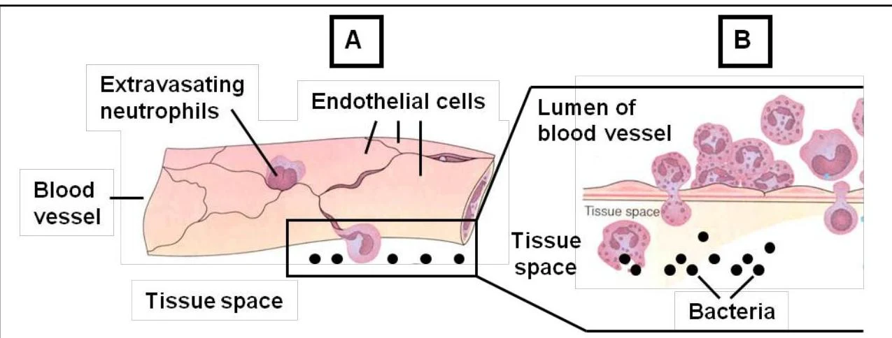
During inflammation, leukocytes undergo extravasation, where they leak out of blood vessels (primarily venules) to reach the site of injury. This movement is often guided by chemokines—a family of cytokines that attract cells to a site via a process called chemotaxis.
Diagram illustrating extravasation of neutrophils from a blood vessel into tissue space, where they interact with bacteria.Figure 4. Recruitment of neutrophils into the inflamed site.
Adaptive Immunity: Specificity and Memory
If innate mechanisms fail, the adaptive immune system is activated. This system is characterized by exquisite specificity for distinct epitopes and the ability to develop memory.
The adaptive response is divided into two types:
Humoral Immunity: Mediated by B lymphocytes (B cells), which produce soluble immunoglobulins (antibodies) that neutralize extracellular pathogens.
Cell-Mediated Immunity: Mediated by T lymphocytes (T cells). These include Helper T cells (CD4+), which secrete cytokines to coordinate the response, and Cytotoxic T cells (CD8+), which kill infected host cells.
Exam Tip: Antigen Recognition Differences
Remember that B cells can recognize antigens in their native form (proteins, lipids, or carbohydrates) via their B cell receptors (BCRs). However, T cells can only recognize protein antigens, and only when they are presented as small peptides on Major Histocompatibility Complex (MHC) molecules by an APC.
The Paradigm of Immunity: Clonal Selection Theory
The Clonal Selection Theory explains how the body manages such vast diversity. The theory posits that:
The body contains billions of unique lymphocytes, each with a unique receptor for a single epitope.
When a specific antigen enters the body, it "selects" the lymphocyte with the matching receptor.
That selected lymphocyte undergoes clonal expansion (rapid proliferation), creating a large army of identical cells.
These cells differentiate into effector cells (to fight the current infection) or memory cells (to provide rapid protection if the antigen returns).
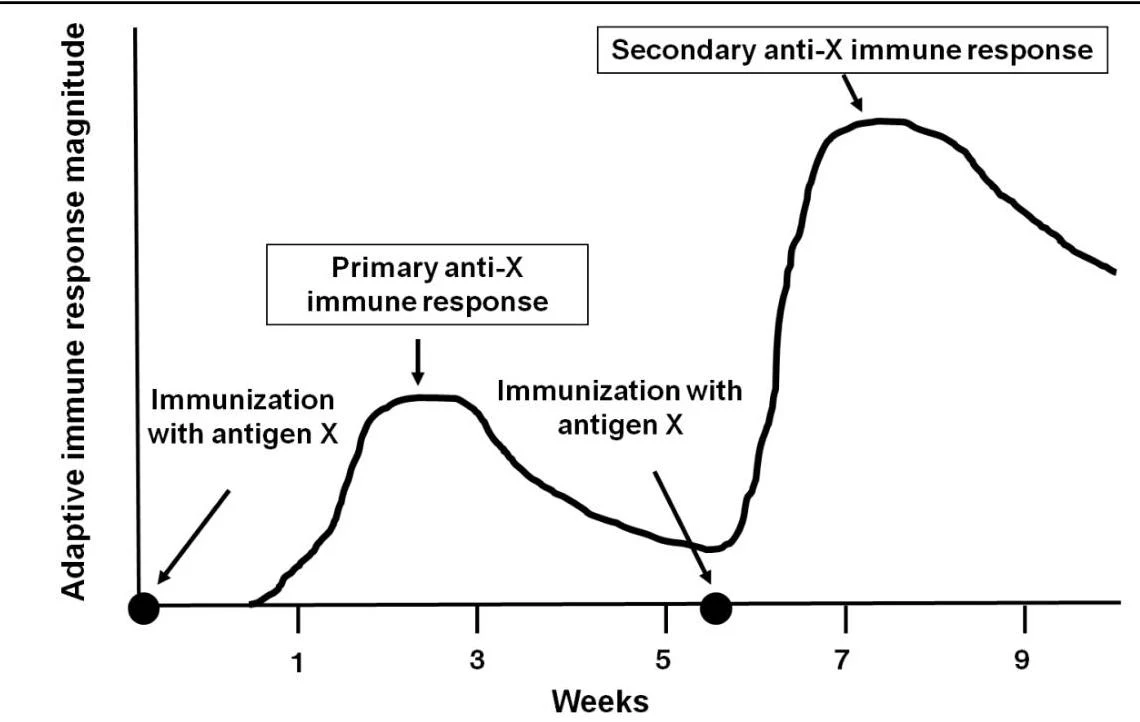
This explains why the secondary immune response (the response upon re-exposure) is much faster and more vigorous than the primary response.
This graph illustrates the primary and secondary immune responses to an antigen over time, showing the adaptive immune response magnitude in weeks.Figure 6. Secondary adaptive immune responses are due to memory lymphocytes.
Secondary Lymphoid Organs
Because lymphocytes are constantly recirculating through the body, they need specialized "meeting places" to encounter antigens. These are known as secondary lymphoid organs.
Lymph Nodes: Filter antigens from the lymph.
Spleen: Filters antigens from the blood. It contains red pulp (for filtering blood and old red cells) and white pulp (where lymphocytes and APCs reside).
MALT (Mucosa-Associated Lymphoid Tissue): Protects mucosal surfaces like the gut (GALT), lungs (BALT), and nose (NALT). In the gut, specialized M cells help transport antigens from the intestinal lumen to the underlying lymphoid tissue.
Diagram of the spleen's internal structure, showing the red pulp and white pulp with its components like the germinal center, primary follicle, marginal zone, and periarteriolar lymphoid sheath (PALS).Figure 5. A schematic diagram of spleen.
Keep reading for free.
Create an account to finish this reading and make it part of your study routine.