


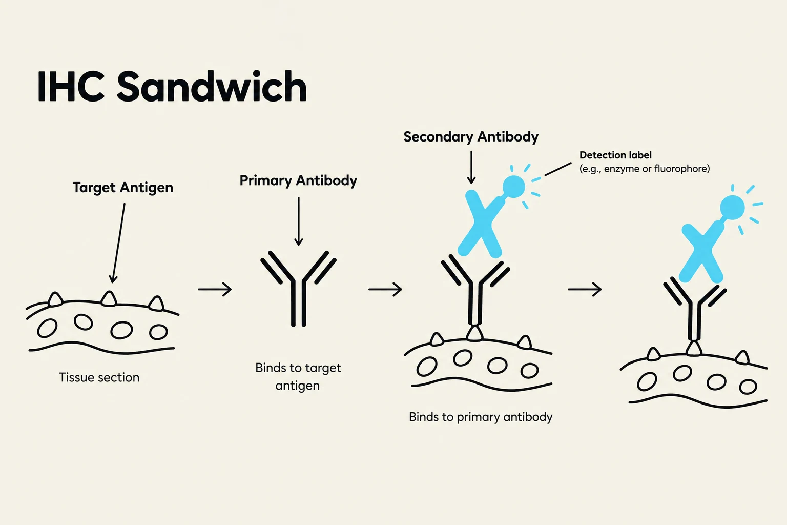
Immunohistochemistry (IHC) is a sophisticated immunoassay used to detect and localize specific proteins, known as biomarkers, within tissue sections. By utilizing the highly specific binding properties of antibodies, IHC allows pathologists to visualize the presence, distribution, and intensity of antigens under a microscope. While it is often categorized as a "special stain," IHC is fundamentally an application of the Enzyme-Linked Immunosorbent Assay (ELISA) principle to intact tissue architecture. This capability provides essential data—ranging from simple binary presence/absence to semi-quantitative or even quantitative protein expression levels—that is critical for cancer diagnosis, classification, and the selection of targeted therapies.
The Total Test Approach and Pre-analytical Factors
In clinical practice, IHC should not be viewed as a single isolated step, but rather as part of a "Total Test" approach. This concept recognizes that the final result is the culmination of a long chain of events, where a failure at any single stage can lead to diagnostic errors, such as false negatives or false positives.
The process begins in the pre-analytical phase, which starts the moment a biopsy is acquired in the surgery room. Key variables include:
- Fixation: Ideally, tissue should be placed in formalin within one hour of removal to prevent autolysis and preserve antigenicity.
- Processing and Embedding: The tissue undergoes dehydration and is embedded in paraffin to create Formalin-Fixed, Paraffin-Embedded (FFPE) tissue.
- Sectioning: The paraffin block is sliced into extremely thin sections, typically around , to allow light to pass through for microscopy.
Exam Tip: Remember that "Standardization starts in the surgery room." If fixation is delayed or improper, even the most expensive antibody cannot recover the lost antigenicity.
Keep reading for free.
Create an account to finish this reading and make it part of your study routine.
Continue reading freeAlready have an account? Log in
Updated