Histology is the microscopic study of cells and tissues, crucial for understanding organ formation, correlating structure to function, and diagnosing diseases. This guide covers principles of microscopy, types of microscopes, their anatomy, and the four basic human tissue types.
Histology is defined as the microscopic study of cells and tissues. By examining the minute details of biological structures, scientists and medical professionals can understand how the specific arrangement of cells forms complex organs and organ systems.
The study of histology is not merely academic; it has vital practical applications in medicine and law:
Functional Correlation: It allows us to correlate the structure of a tissue to its specific biological function and observe how tissues interact with various medicines.
Disease Diagnosis: Histology is essential for diagnosing diseases, such as when a doctor performs a biopsy to analyze tissue samples.
Forensic Science: During autopsies, microscopic examination of tissues can help investigators determine the cause of unexplained deaths.
To view these structures, researchers use a microscope to capture photographs known as micrographs.
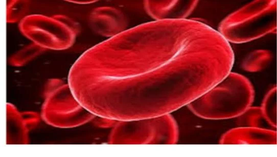
Red blood cells are the most common type of blood cell and the vertebrate's primary means of delivering oxygen to the body tissues—via blood flow through the circulatory system.
Principles of Microscopy
To use a microscope effectively, one must master two fundamental concepts: magnification and resolution.
Magnification
Magnification refers to the enlargement of an object in relation to its actual size. In a compound microscope, the total magnification is calculated by multiplying the magnification of the objective lens by the magnification of the eyepiece. For example, if you are using a 10x eyepiece and a 40x objective lens, the total magnification is:
10×40=400x
Resolution
While magnification makes an image larger, resolution determines how useful that image is. Resolution is the clarity and sharpness of an image, specifically the ability of the microscope to distinguish two closely spaced points as separate entities.
Exam Tip: High magnification does not automatically guarantee high resolution! Even if an image is huge, it is useless if it is blurry and lacks detail.
Several factors can affect the quality of your resolution, including the thickness of the specimen, the quality of the fixation process, and the intensity of the staining used.
Types of Microscopes
Depending on the specimen and the level of detail required, different types of microscopes are employed.
1. Light (Compound) Microscopes
The light microscope is an optical device that uses visible light and a system of lenses to magnify a specimen. The term "compound" refers to the use of two lenses—the objective lens and the eyepiece—working together to produce the final image. These microscopes typically produce 2D images and are staples in histology laboratories.
There are two common modes of light microscopy:
Bright Field Microscopy: This is the most common method, where stained tissue sections are visualized against a bright background.
Dark Field Microscopy: This method is used to observe unstained specimens, such as living cells.
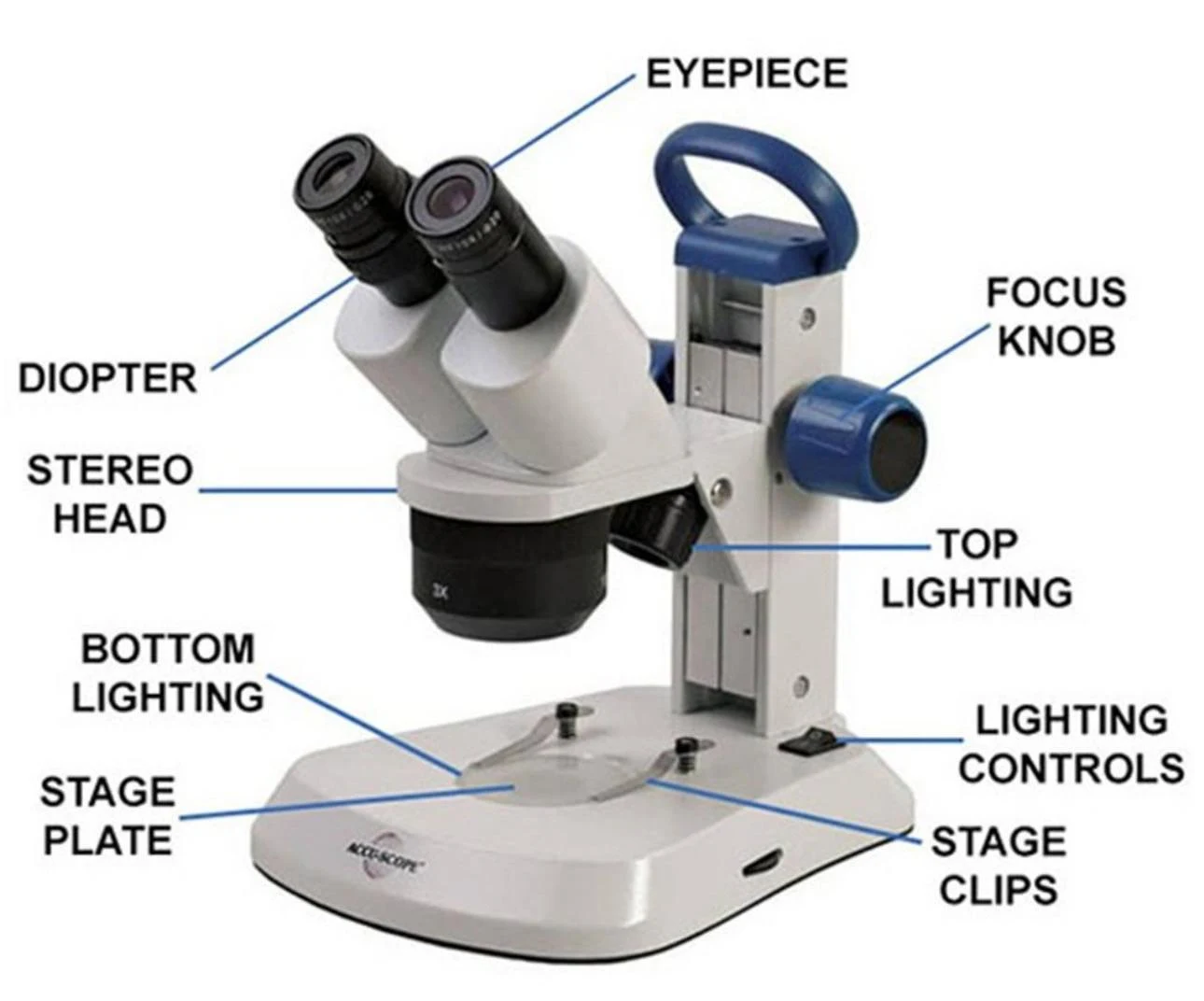
2. Dissecting (Stereo) Microscopes
The dissecting microscope is a variant designed for low-magnification observation. Its primary advantage is that it allows for high-quality, 3D observation of a subject.
Diagram labeling the parts of a stereo microscope, including the eyepiece, diopter, stereo head, focus knob, top lighting, bottom lighting, stage plate, lighting controls, and stage clips.
3. Electron Microscopes
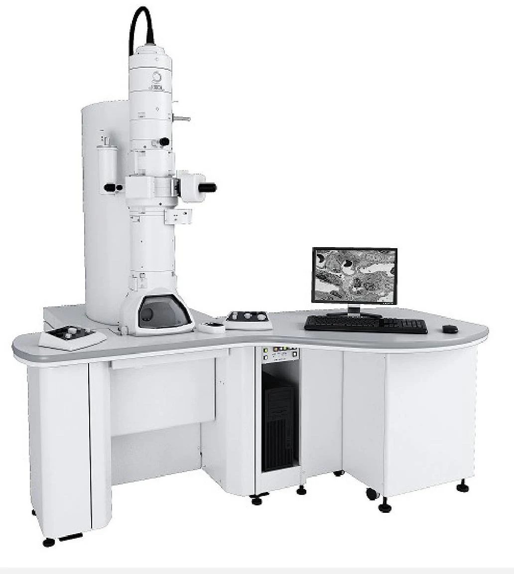
For much higher detail, electron microscopes use a beam of electrons rather than visible light as an illumination source. This technology allows for extreme magnification, capable of viewing biological cells at 20,000x magnification or even higher.
A scanning electron microscope with a computer displaying microscopic images.
Anatomy of a Compound Light Microscope
A compound light microscope is composed of several integrated parts, categorized into optical, mechanical, and adjustment components.
Optical Components
These parts are responsible for the formation and magnification of the image:
Illuminator: The light source, usually a low-voltage bulb.
Condenser: Gathers and focuses the light from the illuminator onto the specimen.
Iris Diaphragm: Adjusts the amount of light that reaches the specimen.
Objective Lenses: These gather light from the object and focus it to create a real image. A standard microscope may have multiple objectives ranging from 4X to 100X.
Eyepiece (Ocular Lens): The lens at the top that the viewer looks through to further magnify the image.
Mechanical and Adjustment Components
These parts provide structure and allow for precise viewing:
Base and Arm: The base supports the microscope, while the arm connects the body tube to the base.
Stage and Stage Clips: The stage is the flat platform where the specimen (mounted on a glass slide) is placed. Stage clips hold the slide in place.
Nosepiece: A rotating turret that holds the objective lenses, allowing the user to switch between different magnifications.
Coarse and Fine Adjustment Knobs: The coarse adjustment is used to bring the specimen into general focus, while the fine adjustment is used to increase detail and sharpness.
Diopter Adjustment: Corrects for vision differences between the user's two eyes.
Safety Note: When using the microscope, always be careful that the objective lens does not touch the glass slide, as this can break the slide and destroy the specimen.
Introduction to Human Tissues
Histology allows us to categorize the human body into four basic types of tissues:
Epithelial Tissue: Provides body coverings and linings.
Connective Tissue: Supports and interlinks other tissues (e.g., bones, tendons).
Muscle Tissue: Contracts to allow for movement and locomotion.
Nervous Tissue: Responsible for fast communication in response to stimuli.
Focus on Epithelial Tissue
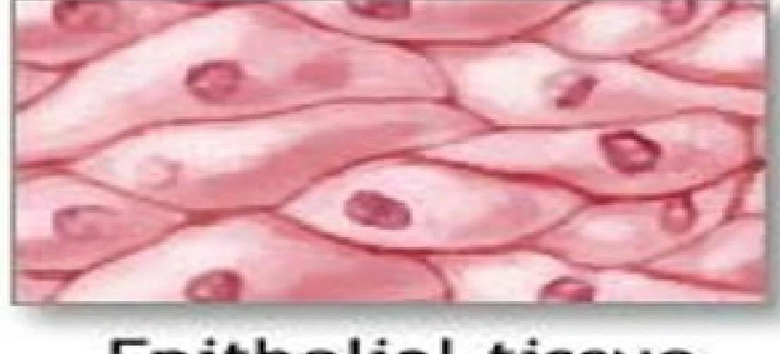
Epithelial tissues are widespread and serve several critical roles, including protection, acting as barriers, secretion, absorption, and permitting the diffusion of substances.
Epithelial tissues possess several unique characteristics:
Cellularity: Cells are in very close contact with little to no intercellular space.
Avascularity: They lack blood vessels; therefore, nutrients must diffuse into the tissue.
Polarity: They have distinct surfaces: an apical surface (top), a basal surface (bottom), and lateral surfaces (sides).
Basement Membrane: They always rest on a basement membrane provided by the underlying connective tissue.
Regeneration: They have a high capacity to produce new cells to replace damaged ones.
Epithelial tissue is a type of biological tissue that lines the surfaces of the body, both internally and externally.
Keep reading for free.
Create an account to finish this reading and make it part of your study routine.