This article explores the journey of human development from germ cells to the establishment of the three primary germ layers and the complex folding that creates the human body plan, with a focus on the early stages relevant to head and neck development.
Embryology can often feel abstract to dental students because the most critical events occur long before a patient is even aware of a pregnancy. However, understanding these early processes is essential for clinical practice. The way the oral cavity, jaws, teeth, salivary glands, and cranial nerves develop is highly coordinated, and many congenital anomalies—such as cleft lip or palate—can be traced back to disruptions in these very first weeks of life.
This article explores the journey from the very first cell to the establishment of the three primary germ layers and the complex folding that creates the human body plan.
From Germ Cells to the Zygote
The biological starting point of human development involves the union of two specialized cells: the male germ cell (sperm) and the female germ cell (oocyte). These cells are produced through a specialized form of cell division called meiosis.
The primary purpose of meiosis is to reduce the chromosome number by half. While a normal human somatic (body) cell contains 46 chromosomes arranged in 23 pairs (the diploid number), germ cells contain only 23 chromosomes (the haploid number). This reduction is vital; if germ cells did not halve their genetic material, the chromosome number would double with every generation.
When fertilization occurs, the sperm and oocyte fuse to form a zygote. This single cell contains the full, restored diploid complement of 46 chromosomes, representing a unique combination of genetic material from both parents.
Early Proliferation: Cleavage, Morula, and Blastocyst
Once the zygote is formed, it begins a period of rapid cell proliferation through a process called mitosis. Unlike meiosis, mitosis is the division of somatic cells that allows for growth and development after fertilization.
During these initial stages, a process known as cleavage occurs. In cleavage, the zygote undergoes repeated mitotic divisions that increase the number of cells, but the overall size of the conceptus does not increase significantly; instead, the individual cells, known as blastomeres, become smaller with each division.
As these cells continue to divide, they form a compact, solid ball of cells called a morula. Eventually, fluid accumulates within this cell mass, transforming the morula into a hollow, fluid-filled structure called a blastocyst.
The blastocyst represents a critical moment of "division of labor" between two distinct cell populations:
Trophoblast: The outer layer of cells that facilitates implantation and the formation of the placenta.
Embryoblast (Inner Cell Mass): The internal cluster of cells that will actually form the embryo proper, including all the tissues of the head and neck.
Exam Tip: Remember the sequence of early development: Zygote → Cleavage → Blastomeres → Morula → Blastocyst. Note that the trophoblast supports the pregnancy, but the embryoblast forms the actual human tissues.
Gastrulation and the Three Germ Layers
Around the second week of development, the embryoblast differentiates into a two-layered structure called a bilaminar disc (consisting of ectoderm and endoderm). However, the most transformative event occurs during week three: gastrulation.
Gastrulation is the process that converts the bilaminar disc into a trilaminar embryonic disc by establishing the three primary germ layers. This process begins with the formation of the primitive streak, a narrow groove on the surface of the embryo. Cells migrate through this streak and reposition themselves to form three distinct layers:
Ectoderm: The outermost layer. It gives rise to the surface epithelium (including oral mucosa and enamel) and the entire nervous system.
Mesoderm: The middle layer. It contributes to muscles, blood vessels, bones, and connective tissues.
Endoderm: The innermost layer. It forms the epithelial lining of the primitive gut and associated structures like the pharynx.
Diagram illustrating the process of gastrulation, showing cell migration through the primitive streak to form the three primary germ layers: ectoderm, mesoderm, and endoderm.
Neurulation and the Importance of Neural Crest Cells
As the germ layers are established, the ectoderm begins to specialize. During weeks three and four, a process called neurulation occurs. The ectoderm overlying the notochord thickens into a neural plate, which then folds to form the neural tube—the precursor to the central nervous system.
A byproduct of this folding is the creation of neural crest cells. These cells are arguably the most important population in craniofacial development. As the neural tube closes, these cells detach and undergo an "epithelial-to-mesenchymal transformation," allowing them to migrate extensively throughout the embryo.
In the head and neck, neural crest cells migrate into the pharyngeal arches and differentiate into a wide variety of tissues, including:
Craniofacial cartilage and bone
Odontoblasts (which produce dentine)
Peripheral nerves and melanocytes
Because these cells are multipotent, they can become many different things depending on their destination. This is why disruptions in neural crest migration can lead to complex syndromes, such as Treacher Collins syndrome, which affects the bones of the face, eyes, and chin.
Embryo Folding and the Primitive Mouth
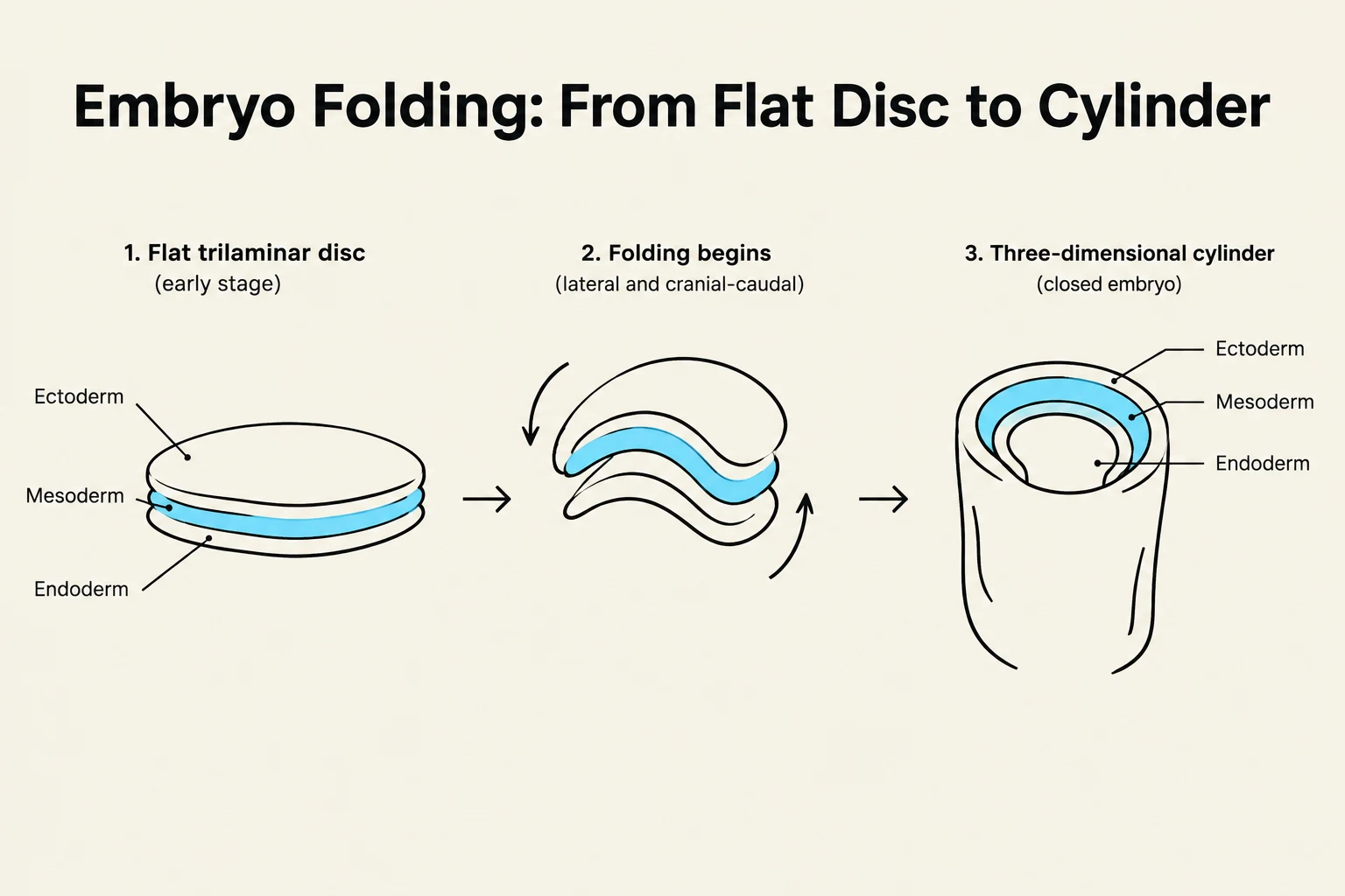
Finally, between weeks three and four, the embryo undergoes embryo folding along two planes: the rostrocaudal (head-to-tail) axis and the lateral axis. This transforms the embryo from a flat disc into a three-dimensional, cylindrical body.
This folding is what creates the stomatodeum, or the primitive oral cavity. Initially, the stomatodeum is separated from the primitive gut (lined by endoderm) by the buccopharyngeal membrane. When this membrane eventually ruptures, it establishes a continuous pathway between the primitive mouth and the pharynx.
Diagram illustrating the process of embryo folding, transforming a flat trilaminar disc into a three-dimensional cylinder, showing the development of ectoderm, mesoderm, and endoderm layers.
Clinical Connection: The complexity of the oral cavity arises from this interface. It is not just an "opening" but a highly coordinated meeting point of surface ectoderm, endoderm-lined gut, and neural crest-derived mesenchyme.
Summary of Developmental Phases
To keep your studies organized, it is helpful to view development in two distinct phases:
Establishment of the basic body plan and germ layers
Phase 2
4–8 Weeks
Morphogenesis
Generation of specific forms (face, jaws, teeth)
Understanding Phase 1 is the essential foundation for understanding how the complex structures of the face and mouth are built in the weeks that follow.
Keep reading for free.
Create an account to finish this reading and make it part of your study routine.