


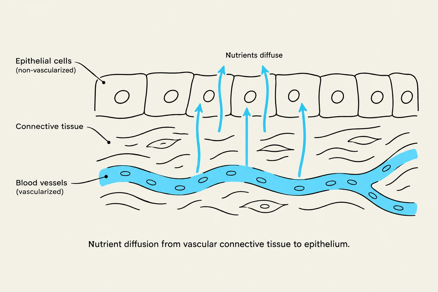
In the study of human anatomy, understanding how the body is constructed requires a deep dive into the different types of tissues that form our organs and systems. Tissues are groups of similar cells working together to perform specific functions. Among the most fundamental are epithelial tissue, which acts as a protective barrier and lining, and connective tissue, which provides the structural framework that holds everything together. While they serve very different roles, they are intimately linked through their biological and nutritional relationships.
Epithelial Tissue: The Body's Lining
Epithelial tissue is characterized by its role as a covering and a lining. It forms the outer surface of the body (the skin) and the inner linings of various organs.
Structural Organization
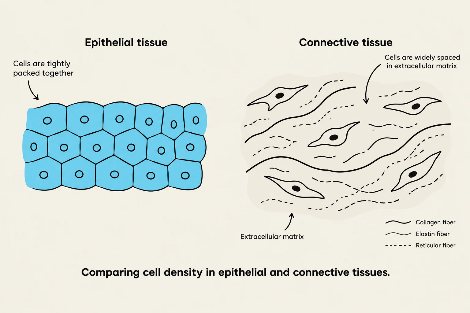
Unlike other tissue types, epithelial cells are tightly packed with virtually no gaps between them. This density is essential for its function as a barrier. The tissue is organized into distinct surfaces:
- Apical Surface: The "top" side of the tissue that faces the exterior of the body or the internal lumen (opening) of an organ. This surface may feature specialized structures like cilia (hair-like projections for movement) or villi (finger-like projections for absorption).
- Basal Membrane: The "bottom" layer that anchors the epithelium to the underlying tissues.
Classification and Examples
Epithelial tissues are classified based on their shape and arrangement. A common type is squamous epithelial tissue, where the cells are flat and resemble fish scales.
The skin, or cutaneous layer, is a primary example of epithelial tissue. The thickness of this layer varies depending on the body part:
Keep reading for free.
Create an account to finish this reading and make it part of your study routine.
Continue reading freeAlready have an account? Log in
Updated