Introduction to Anatomy: Structure and Terminology
An overview of anatomy, covering its scales (gross vs. microscopic), methodologies (regional, systemic, surface), specialized branches, anatomical position, directional terminology, anatomical planes, body movements, and levels of structural organization.
Anatomy is the scientific study of the body's structures and the complex relationships between them. The term itself is derived from the Greek words ana (up) and tome (cutting), reflecting the historical necessity of dissection to uncover the secrets of the human form. By understanding anatomy, we gain insight into how the body is organized, from the microscopic building blocks of life to the massive, integrated systems that sustain existence.
The Scales of Anatomy: Gross vs. Microscopic
Anatomical study is broadly divided into two main scales based on the size of the structures being examined: gross anatomy and microscopic anatomy.
Gross (Macroscopic) Anatomy
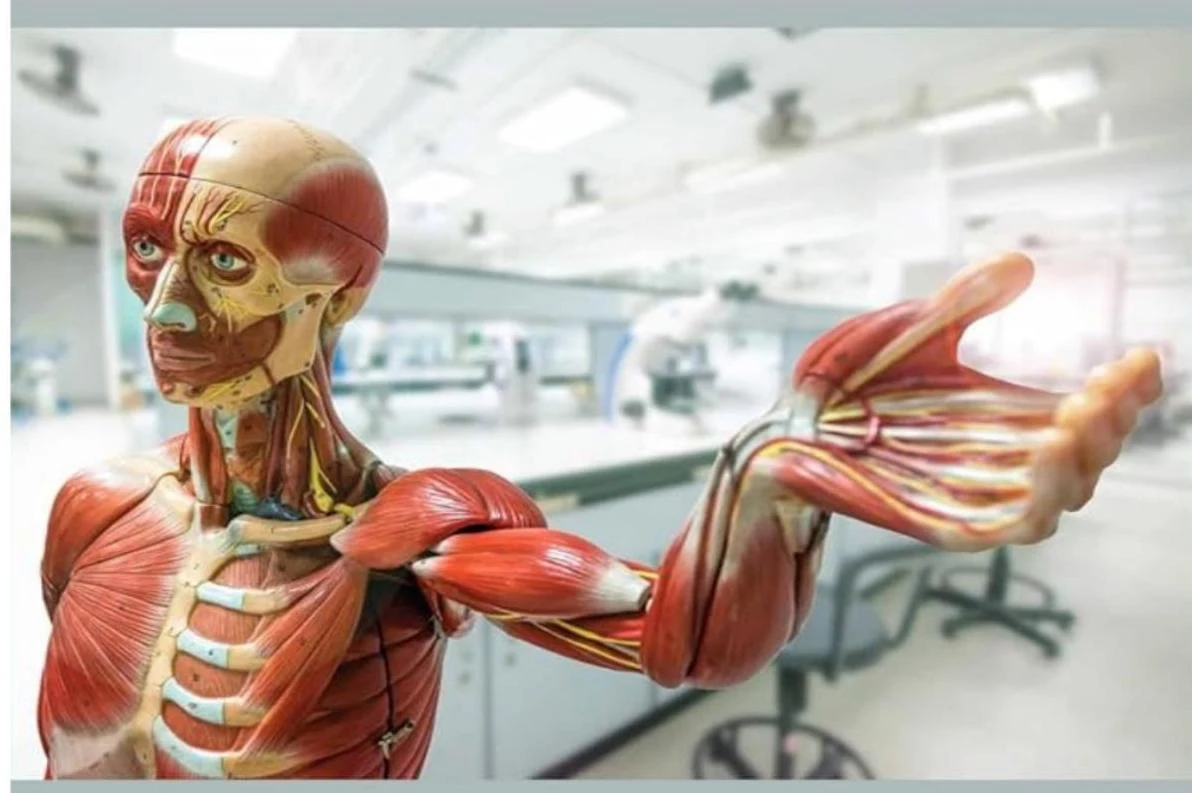
Gross anatomy involves the study of structures that are large enough to be seen with the naked eye. This field is often studied through cadaveric anatomy, which relies on the observation and dissection of deceased bodies to map out organs, bones, and muscles.
Anatomical model of the human muscular system.
Microscopic Anatomy
When structures are too small to be seen without magnification, we enter the realm of microscopic anatomy. This is subdivided into two critical areas:
Cytology: The study of individual cells, focusing on their structure, biochemistry, and behavior.
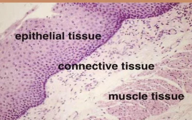
Histology: The study of the composition and structure of tissues, which are groups of cells working together.
Illustration of a microscope, a syringe, and various cells including an animal cell, a plant cell, and bacteria.
Microscopic view of epithelial tissue, connective tissue, and muscle tissue.
Methodologies of Anatomical Study
Depending on the goal of the researcher, gross anatomy can be approached through several different lenses:
Regional Anatomy: This method focuses on specific areas of the body, such as the head, thorax, or limbs, and examines all the various structures (muscles, bones, nerves) within that specific region.
Systemic Anatomy: Instead of looking at a single area, this approach studies the body by grouping organs into functional systems, such as the nervous or circulatory systems, and tracing those systems throughout the entire body.
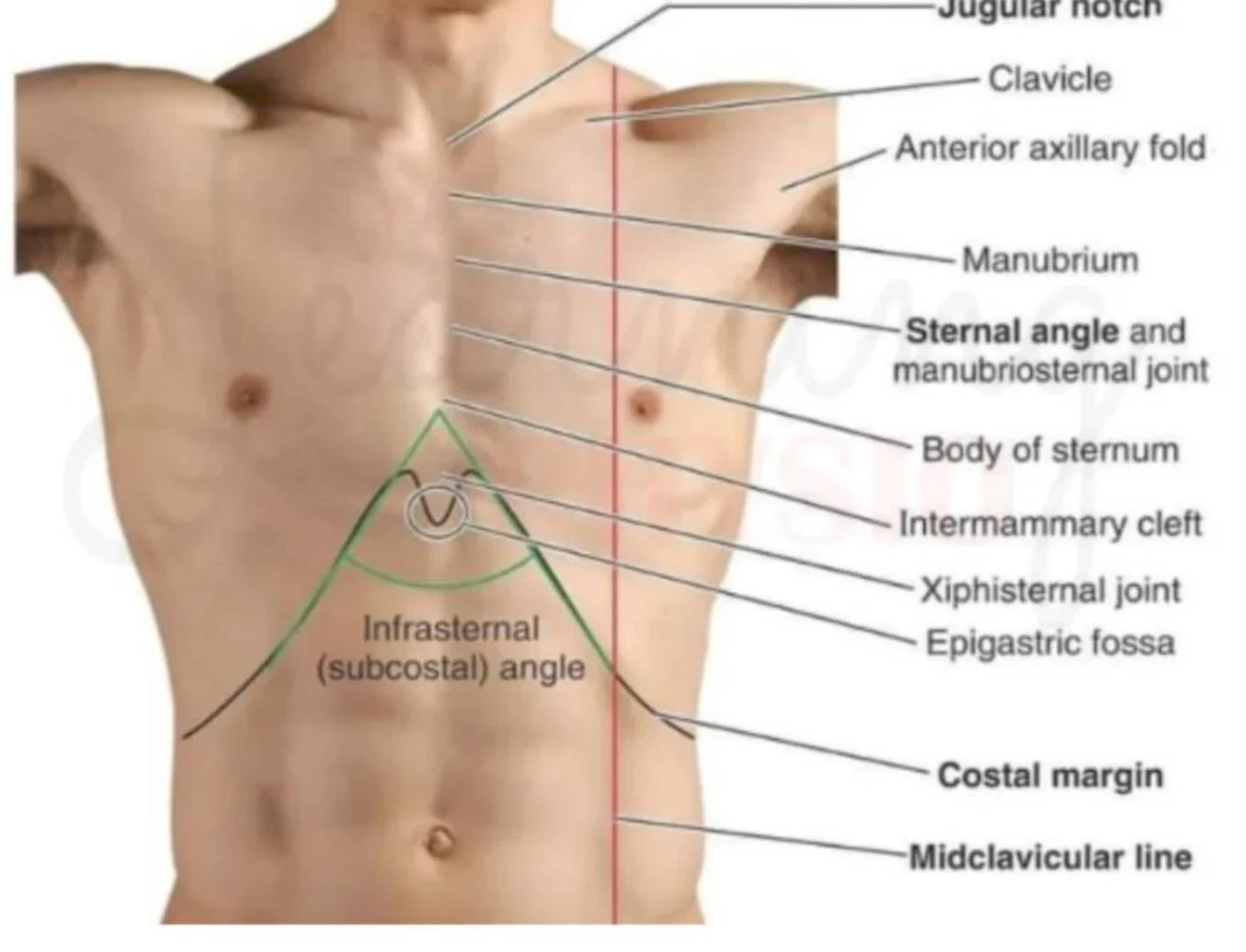
Surface Anatomy: This is the study of visible and palpable features on the body's exterior. By identifying surface landmarks, clinicians can locate deeper underlying structures without needing to perform an incision.
An anatomical illustration of the anterior chest wall, labeling key landmarks such as the jugular notch, clavicle, sternal angle, and costal margin.
Specialized Branches
Beyond the standard classifications, anatomy includes specialized fields that look at the body through different dimensions:
Developmental Anatomy (Embryology): Tracks the structural changes and growth of the body throughout a lifespan, with a specific focus on embryology, which covers development before birth.
Comparative Anatomy: Compares the structures of different species.
Pathological Anatomy: Investigates the structural changes in the body caused by disease.
The Standard: Anatomical Position
To ensure that medical professionals communicate clearly and avoid confusion, all anatomical descriptions are based on a standardized "starting point" known as the anatomical position.
In this position:
The body is standing erect and facing forward.
The head and eyes are directed straight ahead.
The arms are at the sides with the palms facing forward and thumbs pointing away from the body.
The feet are flat, parallel, and pointing forward.
Exam Tip: Remember that "prone" means lying face down, while "supine" means lying face up.
Diagram illustrating anatomical landmarks with directional indicators.
Directional Terminology and Planes
Because the body is a three-dimensional object, we use specific terms to describe where one structure is located in relation to another.
Laterality and Symmetry
Bilateral: Structures on both sides of the body (e.g., kidneys).
Unilateral: A structure on only one side (e.g., the spleen).
Ipsilateral: On the same side of the body.
Contralateral: On the opposite side of the body.
Relative Positioning
Medial vs. Lateral: Medial is toward the midline (middle) of the body; lateral is farther from the midline.
Superior vs. Inferior: Superior is toward the head (above); inferior is toward the feet (below).
Proximal vs. Distal: Used primarily for limbs. Proximal is closer to the trunk or point of origin; distal is farther away.
Anterior (Ventral) vs. Posterior (Dorsal): Anterior is toward the front; posterior is toward the back.
Anatomical Planes
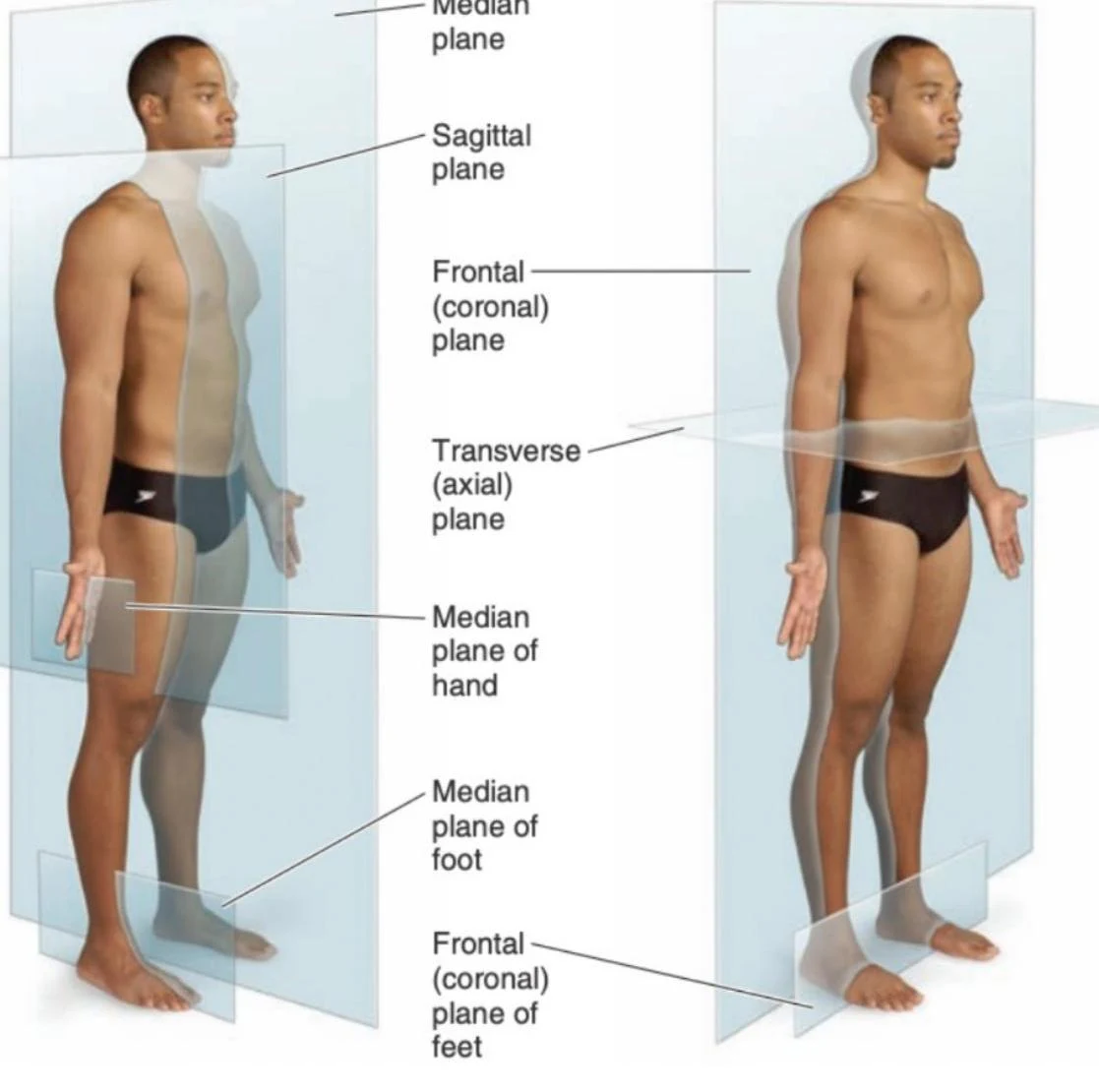
To visualize the body in sections, we use imaginary planes:
Median (Sagittal) Plane: A vertical plane dividing the body into right and left halves.
Frontal (Coronal) Plane: A vertical plane dividing the body into anterior (front) and posterior (back) portions.
Transverse (Axial) Plane: A horizontal plane dividing the body into superior (upper) and inferior (lower) parts.
An illustration showing the anatomical planes of the human body: sagittal, coronal (frontal), and transverse (axial) planes, along with median planes of the hand and foot.
Body Movements
Anatomy also describes how the body moves through space. These movements are often categorized by how they change the angles between bones or their position relative to the midline.
Flexion and Extension: Flexion decreases the angle between two body parts (e.g., bending the elbow), while extension increases that angle.
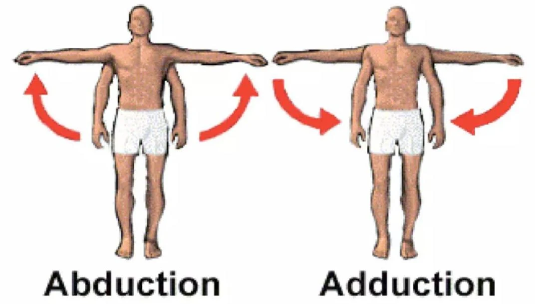
Abduction and Adduction: Abduction moves a limb away from the midline; adduction moves it toward the midline.
Rotation: Turning a structure around its own axis (e.g., internal or external rotation of the hip).
Circumduction: A circular, tri-planar movement.
Pronation and Supination: Specifically used for the forearm; pronation turns the palm down, while supination turns the palm up.
Inversion and Eversion: Movements of the foot; inversion turns the sole inward, while eversion turns it outward.
Diagram illustrating abduction and adduction movements of the arms.
Levels of Structural Organization
To understand how the whole organism functions, we must look at the hierarchy of life. The human body is organized into six distinct levels that build upon one another to maintain homeostasis (biological balance):
Level of Organization
Explanation
Example
Atomic
The smallest unit of an element
Carbon, Oxygen
Molecular
Atoms combining to form complex structures
DNA, Water
Cellular
The smallest unit of life
Muscle cell, Neuron
Tissue
Groups of similar cells with a common function
Muscle, Epithelial
Organ
Two or more tissue types working together
Heart, Stomach
Organ System
A group of organs performing generalized functions
Digestive System
Organismal
All organ systems functioning together
The Human
Homeostasis is the biological process of maintaining a stable internal environment, illustrated by a balance scale representing equilibrium.
By studying these levels, from the chemical to the organismal, we can understand how the complex machinery of the human body operates as a single, unified whole.
Keep reading for free.
Create an account to finish this reading and make it part of your study routine.