A foundational guide to human anatomy and physiology, covering basic chemistry, cells, tissues, and organ systems. It emphasizes a science-based approach, the scientific method, and the concept of homeostasis.
Understanding the human body requires a dual perspective: anatomy, the study of structure, and physiology, the study of function. To master these disciplines, one must look beyond simple memorization and instead adopt a science-based approach that connects the smallest chemical building blocks to the complex, integrated movements of entire organ systems.
The Scientific Foundation: Method and Homeostasis
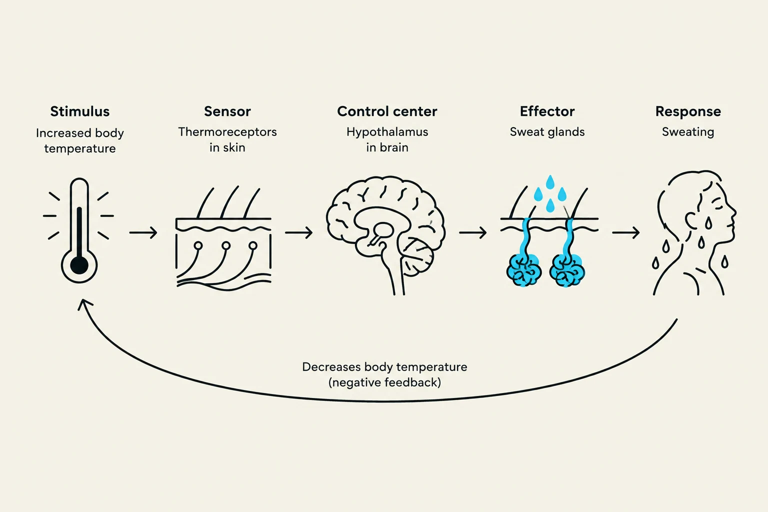
This diagram illustrates the physiological process of thermoregulation, specifically how the body responds to increased temperature through sweating. It shows the stimulus (increased body temperature), sensor (thermoreceptors in the skin), control center (hypothalamus in the brain), effector (sweat glands), and response (sweating), which ultimately decreases body temperature via negative feedback.
Before diving into specific organs, it is essential to understand how biological science operates. The study of anatomy and physiology relies heavily on the scientific method. By applying this method, researchers and students can investigate biological variables, observe patterns, and predict how specific changes might affect bodily functions.
At the heart of all physiological processes is the concept of homeostasis. Homeostasis refers to the sophisticated regulatory mechanisms the body uses to maintain a stable internal environment, even when the external world changes (such as temperature fluctuations or nutrient availability).
Clinical Connection: Homeostatic Dysfunction
In a medical context, many diseases are understood as "homeostatic dysfunctions." This occurs when the body's regulatory mechanisms fail to maintain stability, leading to physiological imbalances that can result in illness or death.
Anatomical Foundations and Terminology
To communicate effectively in a clinical or scientific setting, one must master anatomical terms and identify specific anatomical landmarks. This professional language allows for precise descriptions of location, direction, and planes of the body.
The human body is organized into specific regions and spaces:
Body Cavities and Membranes: The body contains various internal spaces (cavities) protected by specialized membranes that support and separate organs.
Organ Systems: The body is composed of several integrated systems—such as the skeletal, muscular, and nervous systems—that work together to sustain life.
Exam Tip: Precision Matters
Success in this course requires the ability to not only locate anatomical landmarks on models or diagrams but also to correctly spell them. Professional terminology is a key requirement for effective scientific communication.
The Chemistry of Life
The complexity of the human body begins at the molecular level. Basic chemistry provides the framework for understanding how life functions. Molecules are generally categorized into two groups:
Inorganic Molecules: These include essential substances like water, as well as acids and bases, which are critical for maintaining pH balance.
Organic Molecules: These are carbon-based molecules that serve as the primary building blocks of life:
Carbohydrates: Primary energy sources.
Lipids: Essential for energy storage and cell membrane structure.
Proteins: The "workhorses" of the cell, involved in structure, signaling, and catalysis.
Nucleic Acids: The carriers of genetic information (DNA and RNA).
Cellular and Tissue Biology
The hierarchical levels of biological organization are illustrated, starting from molecules like DNA, progressing to cells, then tissues, and finally organs such as the heart.
Moving from molecules to structures, we encounter the cell, the basic unit of life. Understanding cellular organization is vital to understanding how larger systems function.
The Cell and its Mechanics
The plasma membrane acts as a selective barrier, regulating what enters and exits the cell. Furthermore, cells are not static; they undergo the cell cycle, a process of growth and division that allows for tissue repair and development.
The Four Primary Tissue Types
Cells do not work in isolation; they organize into tissues, which are groups of similar cells working together to perform a specific function. There are four primary types of tissue in the human body:
Tissue Type
Primary Function
Epithelial Tissue
Covers body surfaces, lines cavities, and forms glands.
Connective Tissue
Supports, protects, and binds other tissues together.
Muscular Tissue
Responsible for movement through contraction.
Nervous Tissue
Transmits electrical signals to communicate information.
Systemic Anatomy: Skin and Skeleton
As we progress from tissues to organ systems, we examine how these building blocks form complex structures.
The Integumentary System
The skin and membranes constitute the integumentary system. This system serves as the body's first line of defense, providing protection, temperature regulation, and sensory input.
The Skeletal System
The human skeleton is divided into the axial skeleton (skull, vertebral column, rib cage) and the appendicular skeleton (limbs, shoulder girdle, pelvic girdle).
The skeletal system provides the structural framework for the body. It is divided into two main parts:
Axial Skeleton: The central axis of the body, including the skull, vertebral column, and rib cage.
Appendicular Skeleton: The bones of the limbs (arms and legs) and the girdles that attach them to the axial skeleton.
Joints: The points where bones meet, allowing for various degrees of movement and flexibility.
By integrating these levels of organization—from the chemical properties of a protein to the mechanical movement of a joint—students can develop the critical thinking skills necessary to analyze complex clinical scenarios and understand the truly integrative nature of human biology.
Keep reading for free.
Create an account to finish this reading and make it part of your study routine.