An in-depth exploration of the intrinsic muscles of the hand, their anatomical groups, functions, and structural support systems like the extensor hood and palmar fascia. Includes clinical correlations and surface anatomy.
The human hand is a masterpiece of biological engineering, capable of both crushing power and delicate precision. This versatility is largely due to the distinction between extrinsic and intrinsic muscles. While extrinsic muscles originate in the forearm and provide the bulk of the hand's power, the intrinsic hand muscles originate and insert entirely within the hand itself. These twenty small muscles are the primary drivers of fine motor skills, precise finger movements, and nuanced grip strength.
The Five Anatomical Groups of Intrinsic Muscles
To understand hand function, the intrinsic muscles are categorized into five distinct groups based on their location and movement patterns.
1. Thenar Muscles
Located at the base of the thumb, the thenar eminence is responsible for the thumb's wide range of motion, including abduction, adduction, flexion, and opposition. The key muscles in this group are:
Abductor pollicis brevis
Adductor pollicis
Flexor pollicis brevis
Opponens pollicis
2. Hypothenar Muscles
Located at the base of the fifth finger (the pinky), these muscles control the movements of the ulnar side of the hand. They include:
Abductor digiti minimi
Flexor digiti minimi brevis
Opponens digiti minimi
All muscles in the hypothenar group are innervated by the ulnar nerve.
3. Lumbricals
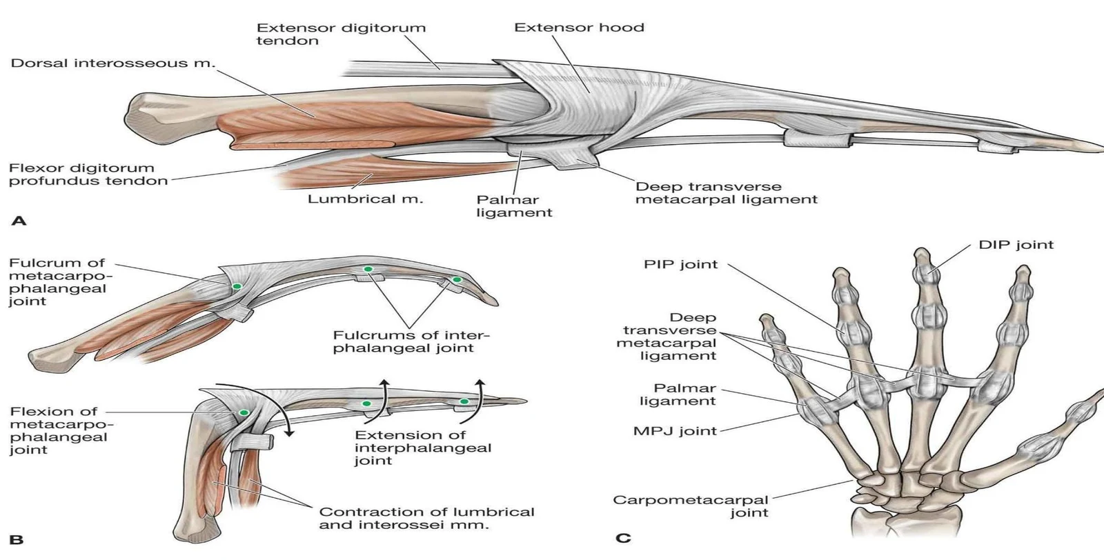
The lumbricals are a unique group of four "worm-like" muscles located in the middle of the palm. Unlike most muscles that connect bone to bone, the lumbricals uniquely originate from the deep flexor tendons (FDP) and insert onto the extensor hood/expansion.
Their primary function is to flex the Metacarpophalangeal joints (MCPJs) while simultaneously straightening (extending) the Proximal Interphalangeal joints (PIPJs) and Distal Interphalangeal joints (DIPJs).
Innervation Pattern:
Lateral two lumbricals: Innervated by the median nerve.
Medial two lumbricals: Innervated by the deep branch of the ulnar nerve.
4. Interossei Muscles
Located between the metacarpal bones, the interossei are essential for fine finger control. They are divided into two functional groups:
Palmar Interossei (PAD): These muscles adduct the fingers (move them toward the middle finger axis). Note that the third digit (middle finger) acts as the central axis and lacks a palmar interossei.
Dorsal Interossei (DAB): These muscles abduct the fingers (spread them apart away from the middle finger axis).
Exam Tip: The Mnemonic
To remember the functions of the interossei, use PAD DAB:
Palmar = ADduct
Dorsal = ABduct
Anatomical illustration of the spaces and bursae in the palm of the hand, including the flexor tendon sheaths, midpalmar space, thenar space, radial bursa, ulnar bursa, and Parona space.
Structural Support: Extensor Hood and Palmar Fascia
The Extensor Hood Compartment
The extensor hood is a flat network of fibrous connective tissue (fibrous aponeurosis) located on the dorsum (back) of each digit. It coordinates finger extension by wrapping around the MCP joint and spreading down the digit. This structure is formed by the extensor digitorum muscle and is reinforced by intrinsic muscles and specific tendons such as the extensor indicis, extensor digiti minimi, and extensor pollicis brevis.
An illustration of the anatomy of the hand, detailing the tendons, ligaments, and joints involved in finger movement. Figure A shows the extensor and flexor tendons and associated muscles. Figure B illustrates the fulcrums and movements of the metacarpophalangeal and interphalangeal joints. Figure C displays the skeletal structure of the hand with labels for various joints and ligaments.
Palmar Fascia
The palmar fascia is a strong, triangular thickening of the deep fascia in the center of the palm. It serves several critical roles: anchoring the skin, improving grip strength, and protecting the underlying tendons and nerves. It is composed of:
Longitudinal fibers: Pretendinous bands extending into the digits.
Transverse fibers: Including the superficial transverse palmar ligament and the natatory ligament (which crosses the web spaces).
Vertical fibers/septa: These connect to metacarpal ligaments to compartmentalize the palm.
Clinical Correlation: Dupuytren's Contracture
Dupuytren's contracture is a localized fibroproliferative disorder of the palmar fascia. Because it arises from internal structures native to the hand (like the fascia), it is classified as an intrinsic hand condition.
Key features include the formation of nodules and thick, dense cords of Type 3 collagen within the palm and the ventral surfaces of the digits. Crucially, this disease involves the fascia and does not involve the tendons.
Risk Factors:
Genetics: Most common in those of North European descent.
Demographics: More common in men over the age of 50.
Medical History: Diabetes, epilepsy, smoking, excessive alcohol consumption, and past hand trauma.
Anatomical illustration of the hand showing Dupuytren's contracture symptoms and relevant anatomical structures including the transverse metacarpal ligament, pretendinous band, palmar aponeurosis, and palmaris longus muscle.
Surface Anatomy and Skeletal Foundation
The Anatomical Snuffbox
The anatomical snuffbox is a triangular depression located on the radial (thumb) side of the dorsum of the hand, which becomes visible when the thumb is extended.
Medial (Ulnar) Border: Tendon of the extensor pollicis longus.
Lateral (Radial) Border: Tendons of the extensor pollicis brevis and the abductor pollicis longus.
Proximal Boundary: The styloid process of the radius.
Floor/Base: The scaphoid and trapezium bones.
Roof: Skin and superficial fascia.
Contents: The radial artery, superficial branch of the radial nerve, and the cephalic vein.
The Carpus
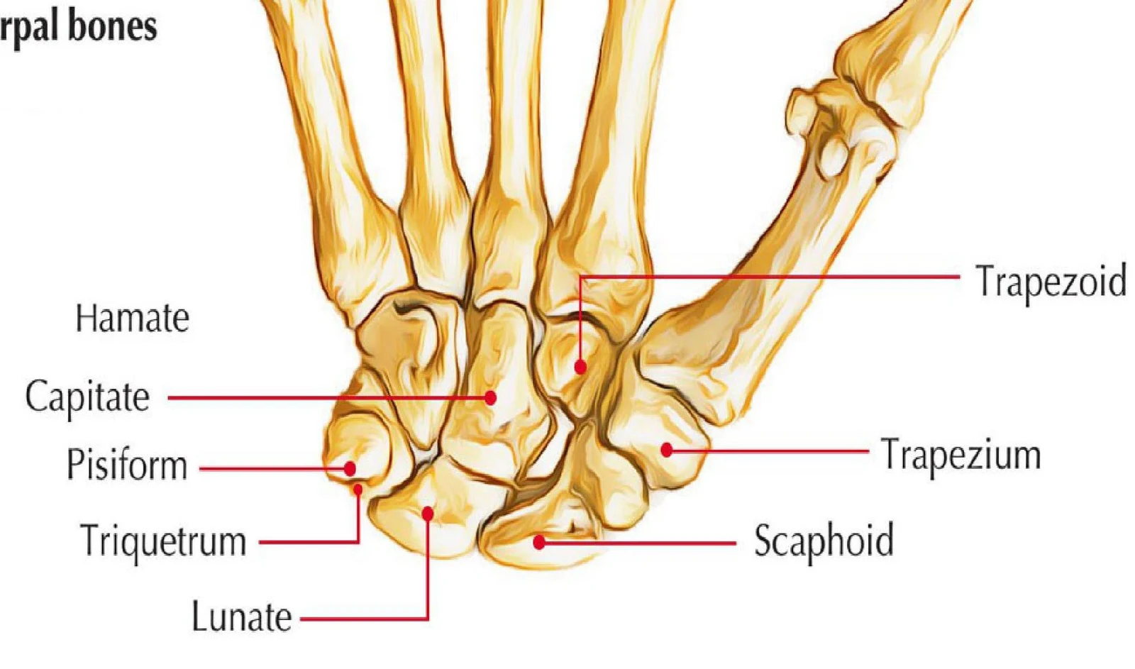
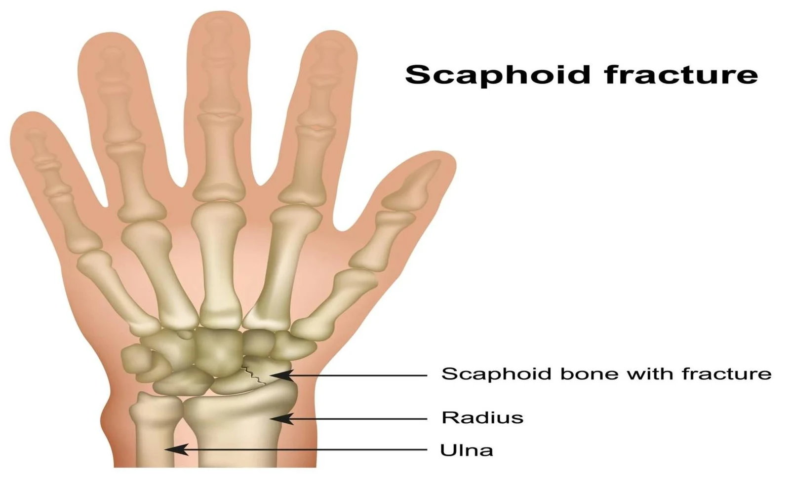
The structural foundation of the wrist is provided by the eight carpal bones, which form the skeletal framework of the carpus. Understanding the arrangement of these bones is vital, particularly when assessing injuries such as fractures of the scaphoid, which forms part of the floor of the anatomical snuffbox.
An illustration of the carpal bones of the wrist, labeling the Hamate, Capitate, Pisiform, Triquetrum, Lunate, Trapezoid, Trapezium, and Scaphoid bones.
An illustration showing a scaphoid fracture in the wrist bones, labeling the scaphoid bone, radius, and ulna.
Keep reading for free.
Create an account to finish this reading and make it part of your study routine.