Intravenous (IV) therapy is a fundamental clinical skill used to maintain homeostasis by delivering fluids, electrolytes, medications, or blood products directly into a patient's vascular system. Because this therapy bypasses the body's natural protective barriers like the gastrointestinal tract, it requires precise regulation of flow rates and vigilant monitoring for complications to ensure patient safety.
Vascular Access Devices
Selecting the appropriate device depends on the duration of therapy and the type of medication being administered.
Peripheral Catheters: These are used for short-term therapy. They are inserted into smaller veins, typically in the hand or arm.
Central Venous Catheters (Central Lines): These are used for long-term access or for delivering irritating medications. They terminate in a large vein near the heart.
PICC Lines (Peripherally Inserted Central Catheters): A type of long-term central access inserted through a peripheral vein in the arm.
Implanted Ports: These are placed under the skin for long-term, intermittent access.
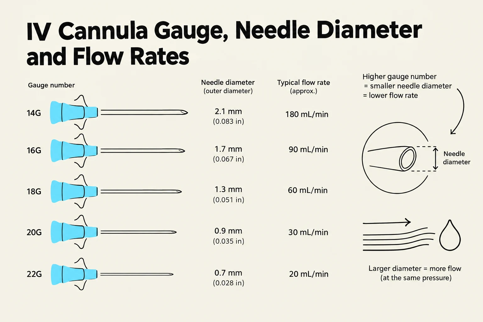
When initiating a peripheral IV, clinicians must select the correct cannula gauge. The rule of thumb is that the larger the gauge number, the smaller the needle diameter.
This chart illustrates the relationship between IV cannula gauge number, needle diameter, and typical flow rates. Higher gauge numbers correspond to smaller needle diameters and lower flow rates, while larger diameters allow for greater flow at the same pressure.
IV Solution Tonicity
Tonicity describes how a solution affects the movement of water between the intravascular space and the cells. Understanding this is critical for preventing cellular damage.
Isotonic Solutions
Isotonic solutions have the same concentration as blood, meaning there is no net movement of water. These solutions "stay" in the intravascular space, making them ideal for fluid replacement in cases of dehydration or hypovolemia.
Hypotonic solutions have a lower concentration than blood. This causes water to move into the cells, which can lead to cellular edema.
Examples: 0.45% Sodium Chloride (Half Normal Saline).
Hypertonic Solutions
Hypertonic solutions have a higher concentration than blood. This draws water out of the cells and into the intravascular space. These must be monitored closely as they can cause fluid overload.
Examples: 3% Sodium Chloride, 50% Dextrose in Water (D50W).
Quick Memory Tip:
Isotonic = Stay (Stays in the bloodstream)
Hypotonic = In (Water moves into the cells)
Hypertonic = Out (Water moves out of the cells)
Regulating Infusion Flow Rates
Clinicians use two primary methods to control the speed of an infusion: electronic pumps or gravity.
Electronic Infusion Pumps
The pump is the most accurate method of regulation. It can be programmed in various modes:
mL/hr: The most common mode for maintenance fluids.
mg/kg/hr or mcg/kg/min: Used for critical, weight-based medications like vasoactive drips.
Gravity IV Lines
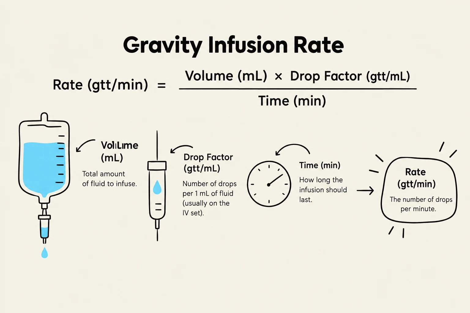
Gravity-fed lines use a roller clamp to regulate flow. To set these, you must calculate the drops per minute (gtt/min) using the drop factor (found on the tubing package).
This diagram illustrates the formula for calculating gravity infusion rate, which is determined by the volume of fluid, the drop factor of the IV set, and the time over which the infusion should occur.
Worked Example:
An order reads: 0.9% NS at 100 mL/hr with a drop factor of 15 gtt/mL.
Flow rate=60100×15=601500=25 gtt/min
Infusion Types and Maintenance
Infusions are categorized by their delivery pattern:
Primary Infusions: Also known as continuous or maintenance infusions (e.g., Normal Saline running steadily).
Secondary Infusions: Often called "piggybacks," these are intermittent infusions used for medications or fluid boluses.
To maintain the system, clinicians must "scrub the hub" (disinfect injection ports) and adhere to strict changing schedules. For example, primary tubing is typically changed every 96 hours, while blood tubing must be changed every 4 hours.
Complications and Interventions
Monitoring the IV site and the patient's systemic response is vital to identify complications early.
Local Complications (At the Site)
Infiltration: Non-vesicant fluid leaks into surrounding tissue. Signs include cool, taut skin and swelling.
Intervention: Stop infusion, discontinue IV, elevate the extremity, and apply a warm compress.
Extravasation: A vesicant (damaging) solution, such as chemotherapy, leaks into the tissue.
Intervention: Stop infusion, discontinue IV, and notify the provider immediately.
Phlebitis: Inflammation of the vein, often feeling "cord-like" or warm.
Intervention: Stop infusion, remove IV, apply a warm compress, and restart at a new site.
Hematoma: A collection of blood in the tissues, often appearing as a hard, painful lump.
Intervention: Elevate the extremity and apply pressure and ice.
Infection: Indicated by redness, warmth, and purulent drainage.
Intervention: Notify the provider and obtain blood cultures as ordered.
Systemic Complications (Whole Body)
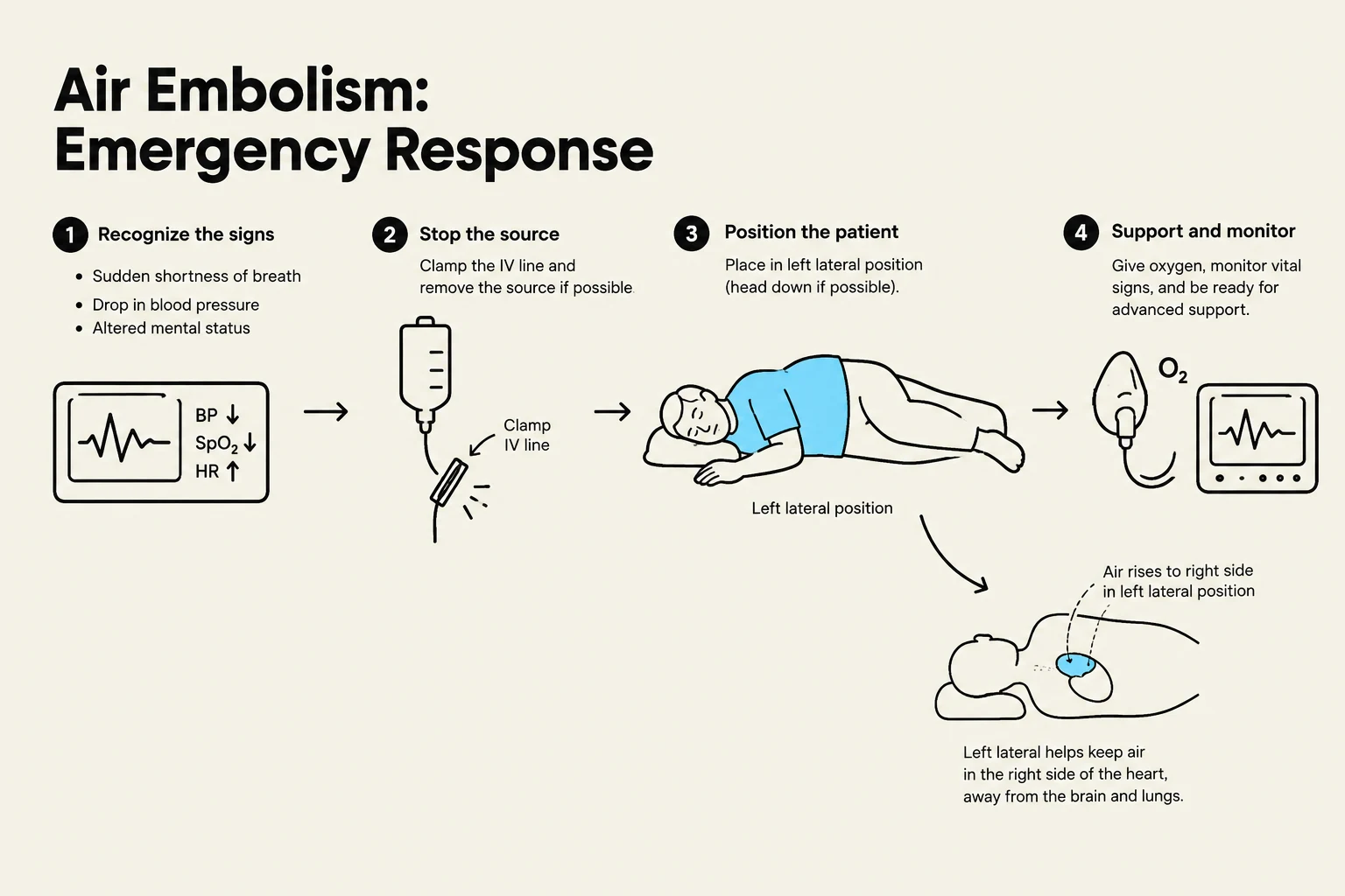
Air Embolism: Air enters the venous system, which can be life-threatening. Signs include sudden dyspnea, chest pain, and tachycardia.
Intervention: Place the patient in left lateral Trendelenburg position, administer 100% oxygen, and notify the provider.
Circulatory Overload: Occurs when fluids are infused too rapidly, leading to hypervolemia. Signs include dyspnea, crackles in the lungs, hypertension, and distended neck veins.
Intervention: Stop the infusion, elevate the head of the bed, assess lung sounds/vitals, and notify the provider.
This diagram illustrates the emergency response steps for an air embolism, including recognizing signs, stopping the source, positioning the patient, and providing support and monitoring.
Discontinuing Peripheral IV Access
Safe removal is necessary to prevent bleeding and infection. After removing the catheter at the same angle of insertion, apply firm pressure with gauze for 2–5 minutes. Always inspect the catheter tip to ensure it is intact and document the patient's tolerance.
Safety First: Always verify the provider's order before starting any infusion and monitor the patient's vital signs and lung sounds throughout the process.
Keep reading for free.
Create an account to finish this reading and make it part of your study routine.