Explains the two main pathways for protein import into cellular organelles: posttranslational import for organelles like mitochondria and cotranslational import for the endomembrane system, detailing the roles of SRP, translocons, and trafficking mechanisms.
In the complex environment of a eukaryotic cell, proteins must be delivered to precise locations to perform their specific biological functions. A protein destined to be a digestive enzyme in a lysosome requires a completely different journey than a protein meant to reside in the mitochondria or be secreted outside the cell. This process of directing proteins to their correct destinations is known as protein sorting, and it relies on two fundamental pathways: posttranslational import and cotranslational import.
The Two Pathways of Protein Import
The destination of a polypeptide determines how it enters its target organelle.
Posttranslational Import
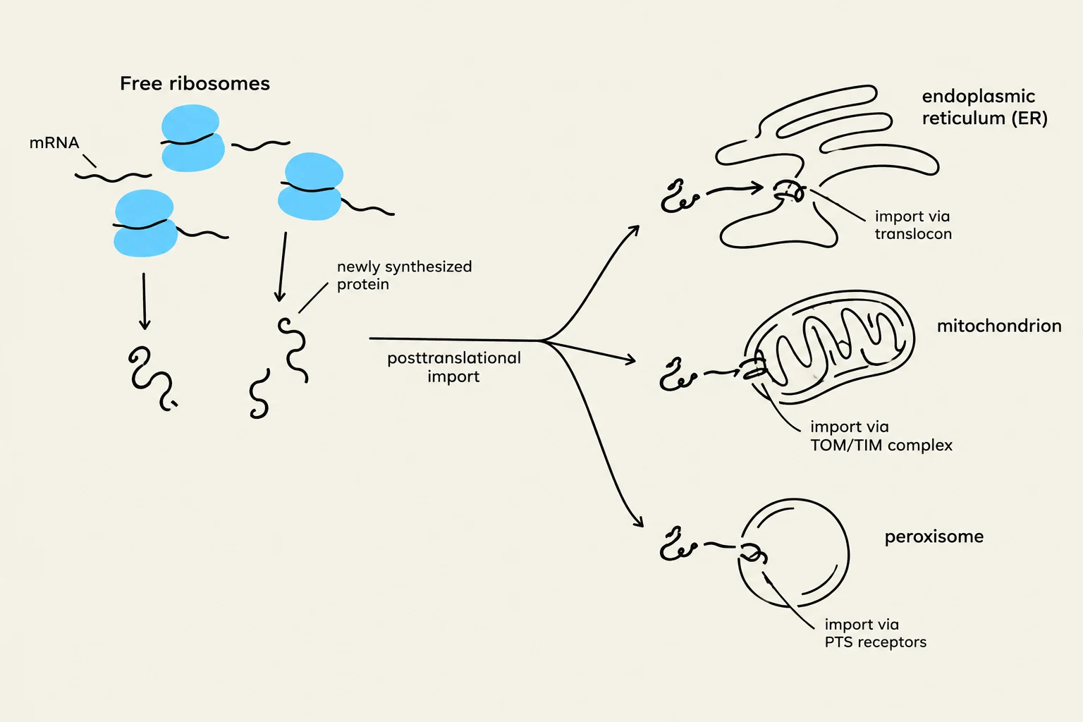
Proteins destined for the cytosol, mitochondria, chloroplasts, peroxisomes, or the nuclear interior undergo posttranslational import. In this pathway, the polypeptide is synthesized entirely on free ribosomes in the cytosol. Once translation is complete and the protein is released from the ribosome, it is recognized by specific targeting signals and taken up by the target organelle.
Cotranslational Import
In contrast, polypeptides destined for the endomembrane system (which includes the endoplasmic reticulum, Golgi apparatus, endosomes, and lysosomes) or for export from the cell utilize cotranslational import. In this mechanism, the ribosome synthesizing the protein attaches to the membrane of the endoplasmic reticulum (ER) while the polypeptide chain is still being elongated. This allows the protein to be inserted into or across the ER membrane simultaneously with its synthesis.
Diagram illustrating the posttranslational import of newly synthesized proteins into various cellular organelles, including the endoplasmic reticulum, mitochondrion, and peroxisome.
The Mechanism of Cotranslational Import
The movement of a protein into the ER is a highly coordinated molecular dance involving several key players. It begins when a ribosome starts translating an mRNA and produces an N-terminal ER signal sequence.
The Role of the SRP and the Translocon
The Signal Recognition Particle (SRP) is the primary mediator of this process. The SRP recognizes and binds to the ER signal sequence as it emerges from the ribosome, which temporarily blocks further translation. The SRP then directs the entire ribosome-mRNA-polypeptide complex to the ER membrane by binding to an SRP receptor.
The complex is then transferred to a translocon, a protein complex that serves as the gateway into the ER. The translocon consists of several essential components:
SRP receptor: Receives the SRP-ribosome complex.
Ribosome receptor: Holds the ribosome in place over the pore.
Pore protein (Sec61 complex): Forms the actual channel through which the polypeptide passes.
Signal peptidase: An enzyme that cleaves the signal sequence from the growing polypeptide once it enters the ER lumen.
As translation resumes, the polypeptide is pushed through the translocon into the ER lumen.
Diagram illustrating the process of protein targeting to the rough endoplasmic reticulum via the signal recognition particle (SRP) pathway.
Trafficking Through the Endomembrane System
Once inside the ER, proteins undergo essential processing, including folding and glycosylation (the addition of carbohydrate chains). From the ER, proteins are routed through the Golgi apparatus via a directional flow of traffic.
Anterograde and Retrograde Trafficking
Protein movement follows two distinct directions:
Anterograde trafficking: The forward movement of materials from the ER to the Golgi (via the cis-Golgi network or CGN), through the medial cisternae, and finally to the trans-Golgi network (TGN). From the TGN, proteins are sent to their final destinations, such as the plasma membrane via secretory vesicles or to lysosomes.
Retrograde trafficking: The backward movement of vesicles from the Golgi back to the ER. This is crucial for maintaining the correct protein composition of the ER and recycling necessary transport machinery.
Exam Tip: Remember that "Anterograde" moves away from the center (toward the cell surface), while "Retrograde" moves back toward the source (the ER).
Maintaining Organelle Identity
Cells use specific molecular "tags" to ensure proteins reach the right place and stay there.
ER Retention and Retrieval
To prevent the loss of essential ER proteins, the cell employs two mechanisms:
Retention tags: Some proteins contain sequences like RXR (Arg-X-Arg) that help keep them within the ER until they are properly assembled.
Retrieval tags: If an ER-resident protein accidentally escapes to the Golgi, it can be "captured" and sent back. These proteins often possess C-terminal sequences like KDEL (in mammals) or HDEL (in yeast).
Sorting in the Golgi
The Golgi apparatus acts as a sophisticated sorting station. Proteins are sorted based on various criteria:
Hydrophobic domain length: Golgi-resident proteins are often sorted by the length of their membrane-spanning domains, which correspond to the increasing thickness of the membranes as one moves from the CGN to the TGN.
Mannose-6-phosphate (M6P) tag: Soluble enzymes destined for the lysosome are specifically tagged with a mannose-6-phosphate group in the Golgi. This tag is recognized by M6P receptors in the TGN, which package the enzymes into vesicles headed for endosomes and lysosomes.
Integral Membrane Proteins
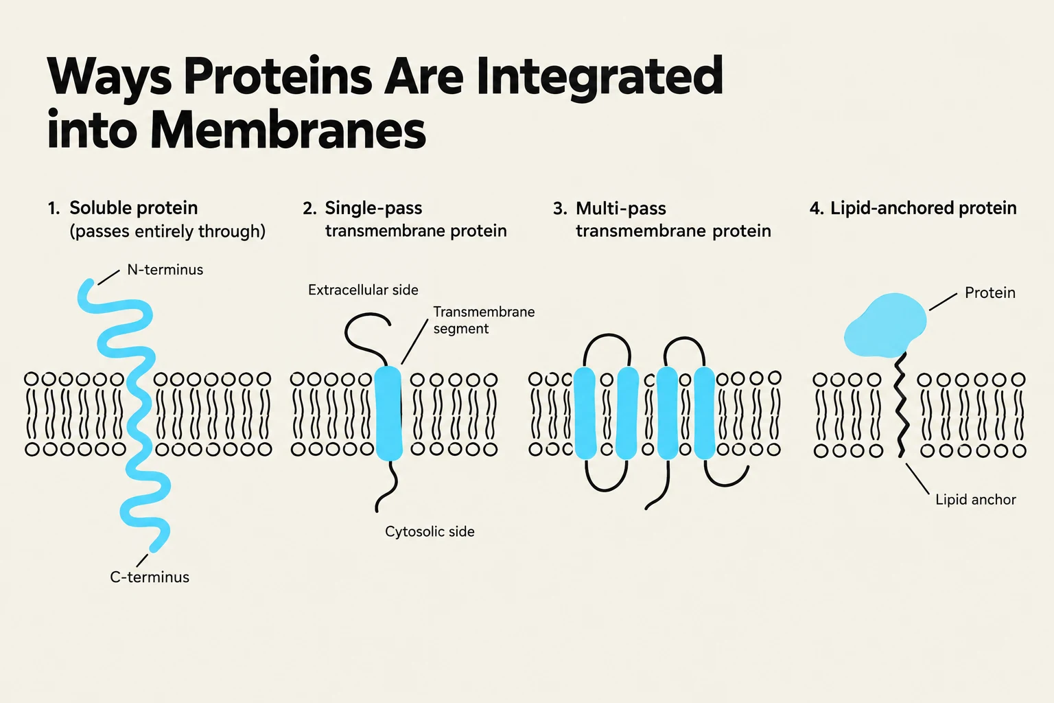
Not all proteins are soluble in the lumen; many must be embedded in the membrane. This is achieved through specialized sequences:
Stop-transfer sequences: These hydrophobic segments halt the translocation of the polypeptide through the translocon, causing the protein to be released laterally into the lipid bilayer.
Start-transfer sequences: These act as internal signals that initiate the insertion of a protein into the membrane during synthesis.
Diagram illustrating four ways proteins are integrated into cell membranes: soluble, single-pass transmembrane, multi-pass transmembrane, and lipid-anchored proteins.
Keep reading for free.
Create an account to finish this reading and make it part of your study routine.