An overview of the integumentary system, common skin disorders like dermatitis and eczema, wound classifications (open and closed), pressure ulcer development, and basic wound care and pain management principles.
The integumentary system is the body's first line of defense against the external environment. It is composed of the skin, hair, nails, and sweat and oil glands. Because the skin serves as a protective barrier, any disruption to its integrity can lead to significant health complications, ranging from minor irritations to life-threatening infections or malignancies. Understanding the various disorders that affect this system and the different classifications of wounds is essential for providing effective patient care.
Common Skin Disorders
Skin disorders can manifest in many ways, affecting the appearance, sensation, and overall health of the patient.
Inflammatory Conditions and Itching
One of the most common issues is dermatitis, which is a general term for skin inflammation. This can be categorized into two main types:
Contact dermatitis: Caused by direct contact with an allergen or an irritating substance.
Irritant dermatitis: A reaction to specific materials such as soaps, detergents, or other chemicals.
Eczema is a specific inflammatory condition characterized by dry, red, and extremely itchy patches of skin, which may sometimes involve papular or vesicular lesions. To manage eczema, clinicians often recommend keeping the skin as moist as possible through the use of lotions or creams.
Closely related to these inflammatory states is pruritus, the medical term for itchy skin. While common in older adults due to dry skin, pruritus can also be caused by pregnancy, allergies, diabetes, or gallbladder and liver diseases. Symptoms may include redness, bumps, blisters, or leathery, scaly skin.
Viral and Malignant Disorders
Beyond inflammation, the integumentary system is susceptible to infections and cancers:
Shingles: A painful viral infection caused by the herpes varicella zoster virus. It can exist in a dormant state and reactivate due to stress, illness, or a weakened immune system, resulting in a painful rash or blisters.
Skin Cancer: A malignant growth that typically develops in the epidermis, making it often easily visible.
Clinical Note on Empathy: Clients experiencing skin disorders may feel embarrassed by physical changes. Always practice empathetic listening and maintain strict confidentiality.
Wound Classifications
A wound is defined as a break in the skin or mucous membrane, which serves as a portal of entry for microbes. Wounds are generally classified into two categories: open and closed.
Open Wounds
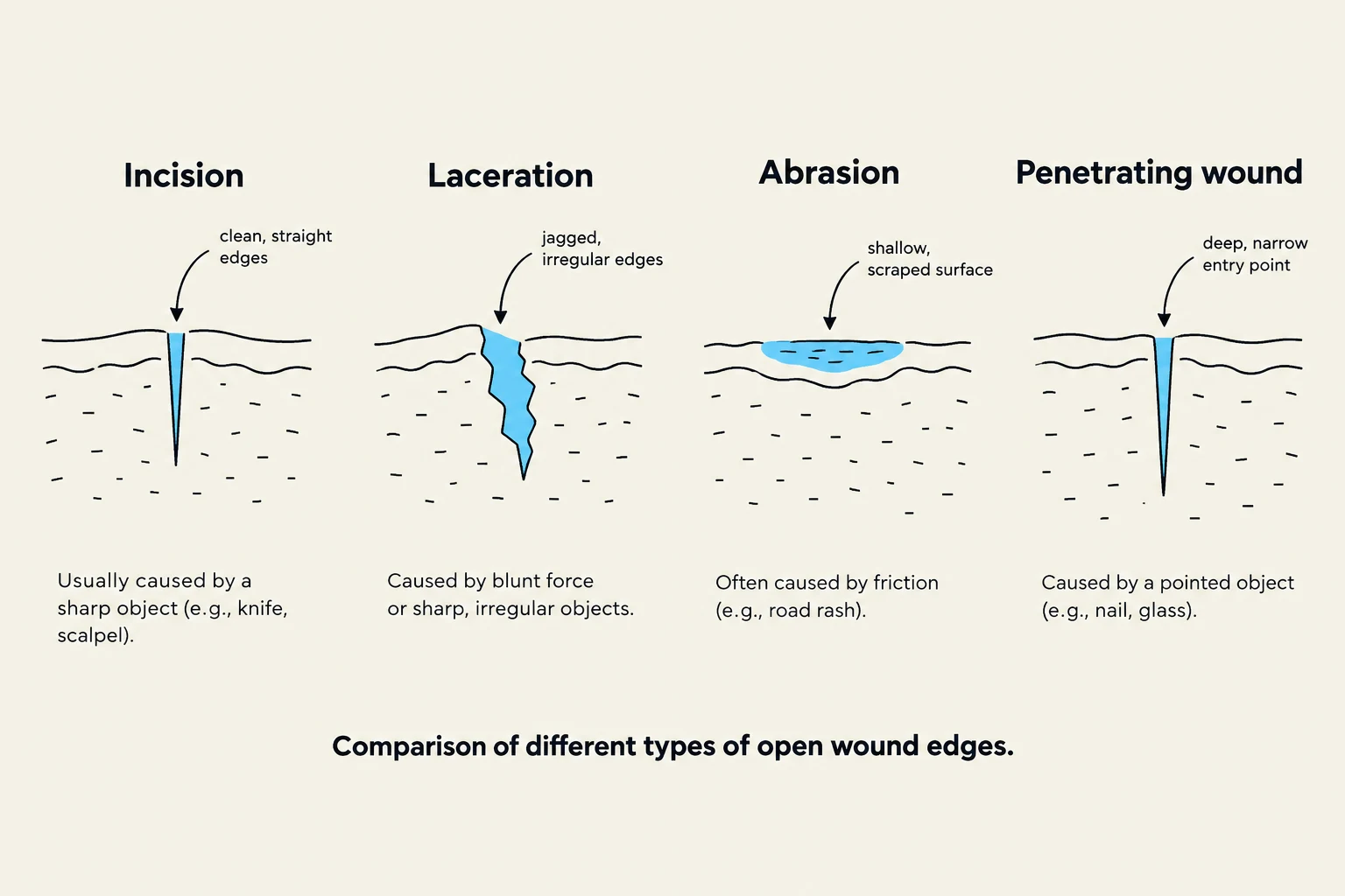
Comparison of different types of open wound edges: Incision, Laceration, Abrasion, and Penetrating wound.
Open wounds involve a break in the skin surface. Common types include:
Abrasion: A partial-thickness wound caused by the skin being scraped or rubbed away.
Incision: A clean, intentional cut made with a sharp instrument, such as during surgery.
Laceration: A wound characterized by torn, jagged edges.
Penetrating Wound: Occurs when skin and underlying tissues are pierced by an object, such as a knife or bullet.
Closed Wounds and Other Injuries
Contusion: A closed wound where tissues are injured (such as a bruise), but the skin remains intact.
Puncture Wound: Caused by sharp objects like nails or animal teeth. While these may exhibit minimal bleeding, they are highly prone to infection and must be treated quickly.
Skin Tear: A specific type of injury where the epidermis separates from the underlying tissue. These are common on the hands, arms, and lower legs of older adults or those with fragile skin, often caused by friction, shearing, or pulling.
Wound Drainage
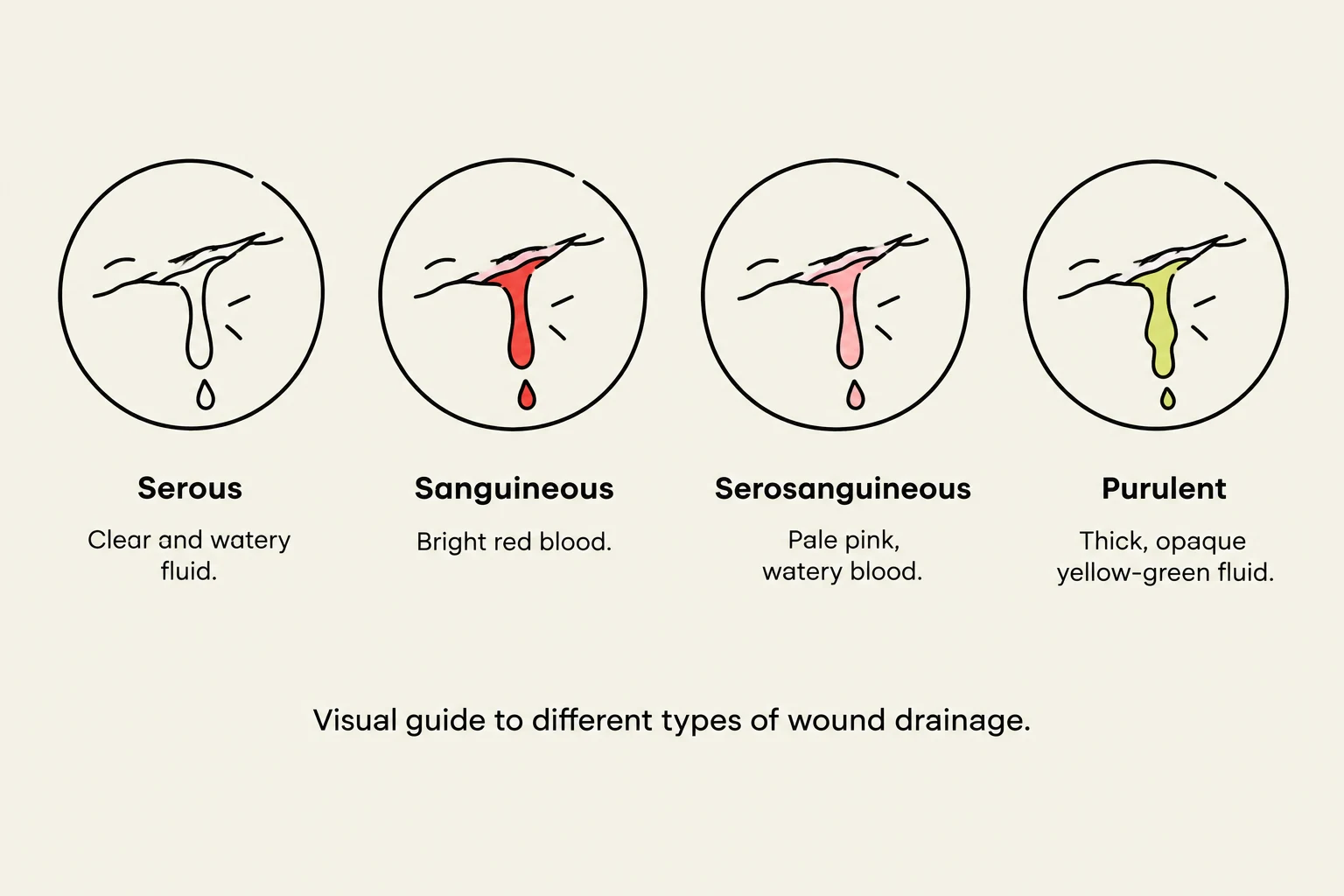
Visual guide to different types of wound drainage: Serous (clear and watery fluid), Sanguineous (bright red blood), Serosanguineous (pale pink, watery blood), and Purulent (thick, opaque yellow-green fluid).
When observing a wound, it is important to note the type of drainage present, as this can indicate the status of an infection:
Serous: Clear, watery fluid.
Sanguineous: Bloody drainage.
Serosanguineous: Thin, watery drainage that is blood-tinged.
Purulent: Thick, green, yellow, or brown drainage that may have a foul odor (often associated with boils or bacterial infection).
Pressure and Tissue Integrity
Pressure-related injuries are a significant concern in healthcare, particularly for immobile clients.
Bony Prominences and Pressure Ulcers
Bony prominences are areas of the body where there is very little fat between the bone and the skin, such as the elbows, heels, hips, and sacrum. When these areas become pressure points—meaning they bear the weight of the body in certain positions—unrelieved pressure can lead to a pressure ulcer (also known as a decubitus ulcer or bedsore).
Contributing factors to pressure ulcers include:
Prolonged pressure and immobility.
Moisture from sweat, urine, or feces.
Friction (rubbing) and shearing (the skin pulling against a surface, such as when a patient slides down in bed).
Poor nutrition, dehydration, and advanced age.
Necrosis and Ulcer Staging
If tissue is deprived of oxygen due to disease or injury, it may undergo necrosis, which is the localized death of body tissue. Necrotic tissue may appear bluish or black.
Pressure ulcers are staged based on the depth of the injury:
Stage 1: Skin is red and does not return to normal color after pressure is removed.
Stage 2: Skin is broken, appearing as cracks, blisters, or peeling.
Stage 3: The ulcer extends into the subcutaneous fat layer.
Stage 4: The ulcer is deep, extending to the muscle or bone.
Wound Care and Pain Management
The primary goals of wound care are to prevent infection, prevent further injury, manage blood loss, and promote client comfort.
Prevention Strategies
For healthcare assistants (HCAs), prevention is the most effective tool. Because pressure ulcers are painful and can be fatal, proactive care is required:
Repositioning: Follow the care plan to turn the client regularly.
Skin Care: Keep skin clean and dry, but avoid rubbing reddened or bony areas.
Mobility: Encourage movement to stimulate circulation.
Nutrition: Encourage adequate hydration and nutrition to support healing.
Assessing and Managing Pain
Pain is a subjective experience; it is "what the person says it is". It can be acute (lasting less than 6 months), persistent (longer than 6 months), referred (felt away from the source), or phantom (felt in a limb that is no longer there).
To assess pain effectively, clinicians often use the PQRST acronym:
P (Provokes/Palliates): What makes it better or worse?
Q (Quality): Is it stinging, dull, sharp, or burning?
R (Region/Radiation): Where is it, and does it move?
S (Severity): On a scale of 1–10, how bad is it?
T (Timing): When did it start?
Exam Tip: Pain Assessment
When reporting pain to a supervisor, be specific. Use the client's exact words to describe the quality of the pain and use the PQRST method to provide a complete picture.
Once pain is reported, HCAs can assist through non-pharmacological interventions such as repositioning, providing a warm blanket, or using distraction techniques like playing soft music.
Keep reading for free.
Create an account to finish this reading and make it part of your study routine.