Innate Immunity: The Body's Second Line of Defense
An overview of innate immunity, detailing the cellular components, phagocytosis, inflammation, the lymphatic system, and fever as the body's non-specific defense mechanisms against pathogens.
When the first line of defense—the physical and chemical barriers like skin and mucous membranes—is breached, the body immediately activates its second line of defense. Unlike the first line, which acts as a preventative wall, the second line is a non-specific, active response designed to destroy invading pathogens and repair damaged tissues. This system relies on a complex interplay of specialized cells, chemical signals, and systemic responses to contain an infection before it can spread.
The Cellular Foundation: Hematopoiesis and Leukocytes
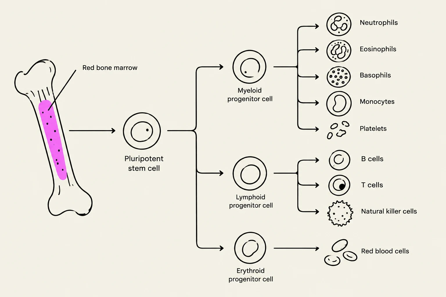
To understand how the body fights infection, we must first look at how its "soldiers" are produced. All immune cells originate from pluripotent stem cells in the red bone marrow through a process called hematopoiesis. These stem cells have the remarkable ability to differentiate into various specialized cell types.
The blood contains several formed elements, including erythrocytes (red blood cells), platelets, and leukocytes (white blood cells). While erythrocytes transport oxygen, leukocytes are the primary actors in the immune response. A standard white blood cell count typically ranges from 5,000 to 10,000 cells per cubic millimeter.
Leukocytes are categorized into several types, each with a specific role:
Neutrophils: The most abundant (60%–70%), acting as rapid responders to bacterial infections.
Lymphocytes: Essential for adaptive immunity (20%–25%).
Monocytes: These differentiate into macrophages (3%–8%).
Eosinophils and Basophils: Involved in allergic responses and parasitic infections.
Diagram illustrating the differentiation of pluripotent stem cells in red bone marrow into various blood cells, including myeloid, lymphoid, and erythroid progenitor cells, which further develop into neutrophils, eosinophils, basophils, monocytes, platelets, B cells, T cells, natural killer cells, and red blood cells.
Phagocytosis: The Primary Attack Mechanism
One of the most critical non-specific responses is phagocytosis, a process where specialized cells known as phagocytes engulf and digest microorganisms. Phagocytes include neutrophils and macrophages. Macrophages can be "fixed" (resident in specific tissues) or "wandering" (roaming the tissues to find infection sites).
The process of phagocytosis occurs in four distinct stages:
Chemotaxis: Chemical signals (such as cytokines) act as a breadcrumb trail, attracting phagocytes to the exact site of the microorganism.
Adherence: The phagocyte physically attaches to the surface of the pathogen.
Ingestion: The cell membrane wraps around the microbe, pulling it inside. This stage is significantly enhanced by opsonization, where serum proteins coat the pathogen, essentially "tagging" it to make it easier for the phagocyte to grip.
Digestion: The ingested microbe is trapped in a vesicle and fused with a lysosome to form a phagolysosome, where enzymes destroy the invader.
Exam Tip: Watch out for "Microbial Evasion" questions! Pathogens have evolved ways to survive this process. For example, Streptococcus pneumoniae uses a capsule to inhibit adherence, while Mycobacterium tuberculosis prevents the fusion of the phagosome and lysosome to stay alive inside the cell.
Inflammation: The Localized Response
When tissue is damaged, the body initiates inflammation to limit the spread of the injurious agent and begin the repair process. Inflammation is characterized by four classic signs: redness (erythema), swelling (edema), heat, and pain.
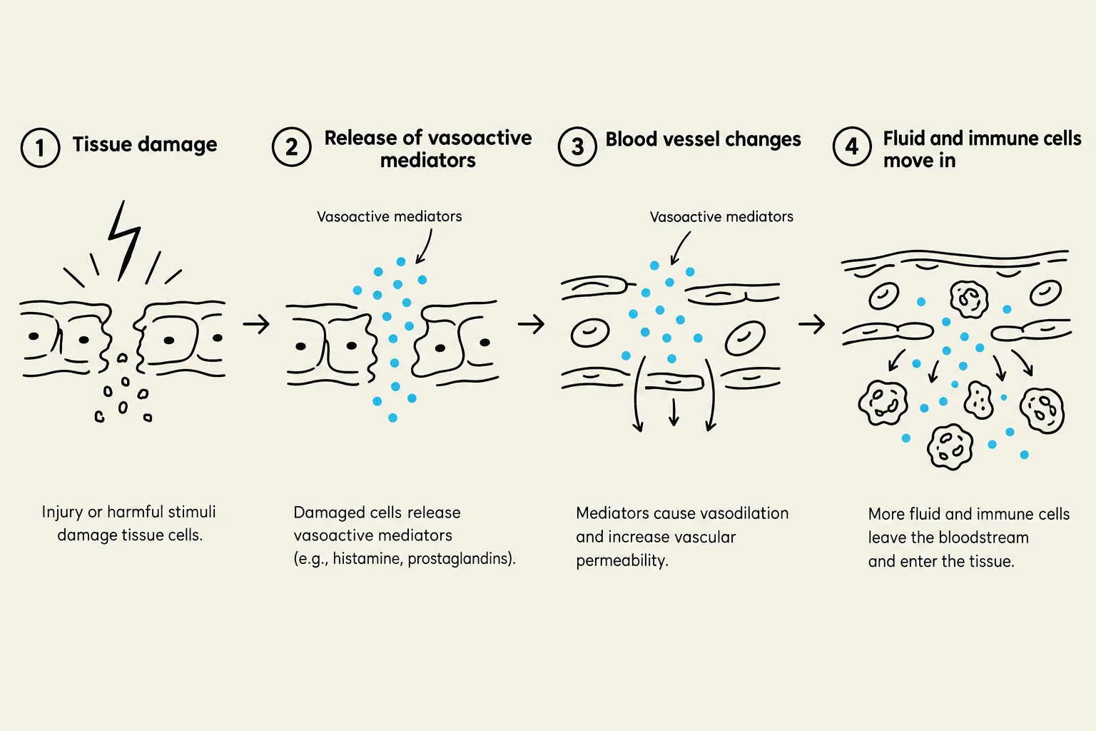
This response is driven by vasoactive mediators—chemical messengers that alter blood vessel behavior. These include:
Histamine: Released by mast cells to increase blood flow.
Kinins, Prostaglandins, and Leukotrienes: Mediate pain and inflammation.
Cytokines: Signal other immune cells to the area.
As these mediators are released, they cause vasodilation (widening of blood vessels) and increased capillary permeability. This allows phagocytes to leave the bloodstream and enter the tissue through a process called phagocyte migration. This involves margination (phagocytes sticking to the vessel walls) and diapedesis (the actual squeezing of the cells between endothelial cells to reach the site of injury).
This diagram illustrates the inflammatory response, showing tissue damage, the release of vasoactive mediators, blood vessel changes like vasodilation and increased permeability, and the subsequent movement of fluid and immune cells into the affected tissue.
The Lymphatic System: The Open Transport Network
While the circulatory system is a closed loop, the lymphatic system is an "open" system that helps manage fluid balance and immune surveillance. It consists of lymph, lymphatic vessels, lymphoid tissue, and red bone marrow.
The system collects interstitial fluid (fluid between cells) and moves it through lymphatic capillaries into larger vessels. This fluid, now called lymph, carries microbes away from the site of infection and transports them to lymph nodes. In the lymph nodes, a high concentration of lymphocytes and macrophages wait to intercept and destroy the pathogens.
Fever: Systemic Defense
If an infection is significant, the body may trigger a fever—an abnormally high body temperature. This is not a failure of the body, but a calculated defense mechanism.
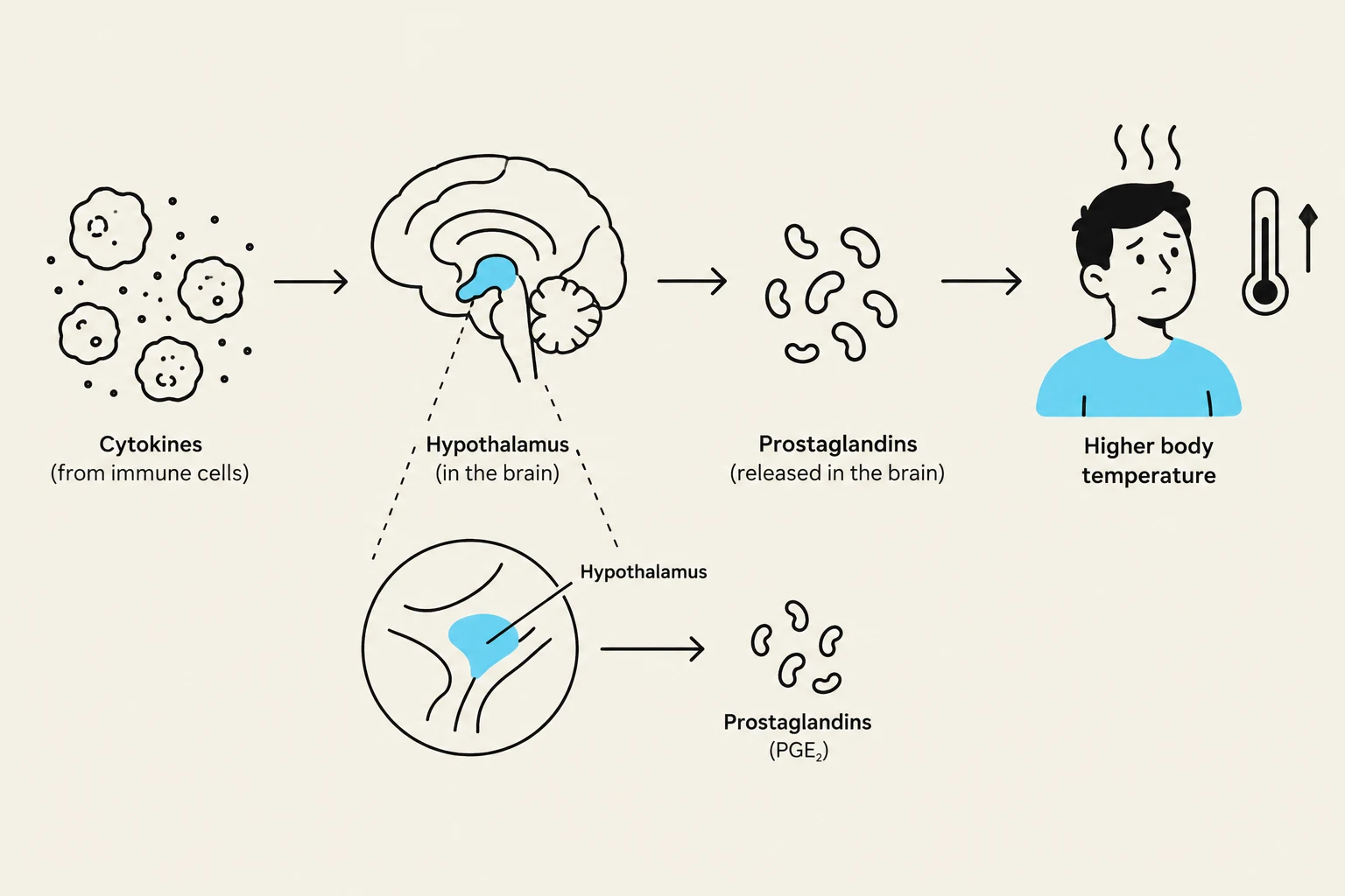
The process begins when cytokines trigger the release of prostaglandins in the hypothalamus, which acts as the body's thermostat. The hypothalamus "resets" to a higher temperature, causing the body to constrict blood vessels and shiver to generate heat.
The primary goal of a fever is to increase the body temperature to a level that slows down the replication rate of microbes, giving the immune system a better chance to win the battle.
This diagram illustrates how cytokines from immune cells trigger the hypothalamus in the brain to release prostaglandins, leading to an increase in body temperature.
Keep reading for free.
Create an account to finish this reading and make it part of your study routine.