An overview of the body's innate immune system, its first and second lines of defense, and the molecular mechanisms involved in recognizing and eliminating pathogens.
When a pathogen enters the human body, it encounters a sophisticated, multi-layered defense system designed to identify and destroy invaders before they can cause widespread damage. This system is broadly divided into two functional categories: innate immunity and adaptive immunity. Understanding the distinction between these two, as well as the specific cellular and molecular mechanisms at play, is fundamental to understanding how the body maintains health and avoids disease.
The Fundamentals: Immunity vs. Susceptibility
To study the immune system, we must first define two opposing concepts: immunity and susceptibility.
Immunity is the body's ability to ward off disease.
Susceptibility represents a lack of resistance to a specific pathogen, making an individual more likely to fall ill.
The body utilizes two distinct strategies to achieve immunity: innate and adaptive.
Innate Immunity
Innate immunity serves as the body's first line of defense. It is nonspecific, meaning it provides a generalized defense against any pathogen rather than targeting a specific strain. Because these defenses are always present from birth, they can respond rapidly to threats, though they require significant energy to maintain.
Adaptive Immunity
In contrast, adaptive immunity is highly specific to a particular pathogen. While it responds more slowly to an initial infection than the innate system, it possesses a memory component. This allows the immune system to "remember" a specific virus or bacteria, enabling a much faster and more effective response upon subsequent exposures.
Exam Tip: If a question asks which system provides "immediate, non-specific" protection, the answer is always innate immunity. If it mentions "memory" or "specificity," it is referring to adaptive immunity.
Molecular Recognition: How the Body "Sees" Invaders
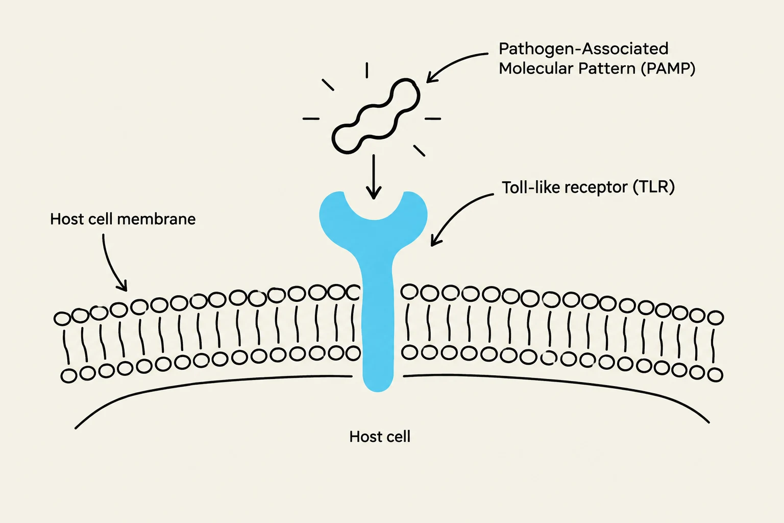
How does the innate immune system know that a cell is a "friend" and not a "foe"? The answer lies in molecular recognition. Host cells use specialized sensors called Toll-like receptors (TLRs) to identify and attach to pathogen-associated molecular patterns (PAMPs).
PAMPs are molecular structures that are common to many pathogens but are not found in human cells. When a TLR binds to a PAMP, it triggers a signaling cascade that induces the release of cytokines. Cytokines are signaling proteins that regulate the intensity and duration of the immune response, essentially acting as the "alarm system" for the body.
Common examples of PAMPs include:
LPS (Lipopolysaccharide): Found in the cell walls of certain bacteria.
PGN (Peptidoglycan): A component of bacterial cell walls.
LTA (Lipotechoic acid): Found in Gram-positive bacteria.
MAN (Mannan): Sugars found on various pathogens.
CpG (CG islands): Specific DNA sequences often found in bacteria.
Diagram illustrating the interaction between a pathogen-associated molecular pattern (PAMP) and a Toll-like receptor (TLR) on a host cell membrane.
Monitoring Health: White Blood Cell Counts
The health and activity of the immune system can be monitored by measuring the concentration of white blood cells (WBCs), also known as leukocytes, in the blood. A normal range for a healthy adult is typically between $5,000$ and $10,000$ WBCs per cubic millimeter (mm3).
Clinicians look at both the total count and the relative percentages of different types of leukocytes to diagnose issues:
Cell Type
Normal Percentage
Primary Role/Notes
Neutrophils
$60% - 70%$
Most abundant; first responders to infection
Lymphocytes
$20% - 25%$
Key players in adaptive immunity
Monocytes
$3% - 8%$
Develop into macrophages
Eosinophils
$2% - 4%$
Involved in parasitic infections
Basophils
$0.5% - 1%$
Involved in allergic responses
Abnormalities in these counts provide vital clues. For example, a high WBC count (leukocytosis) may indicate a bacterial infection or an autoimmune disease, while a low count (leukopenia) might suggest a viral infection or certain cancers.
The Second Line of Defense: Phagocytosis and Inflammation
Once a pathogen bypasses physical barriers like the skin or mucous membranes, it encounters the second line of defense: phagocytes. The term "phagocyte" comes from the Greek phago (to eat) and cyte (cell).
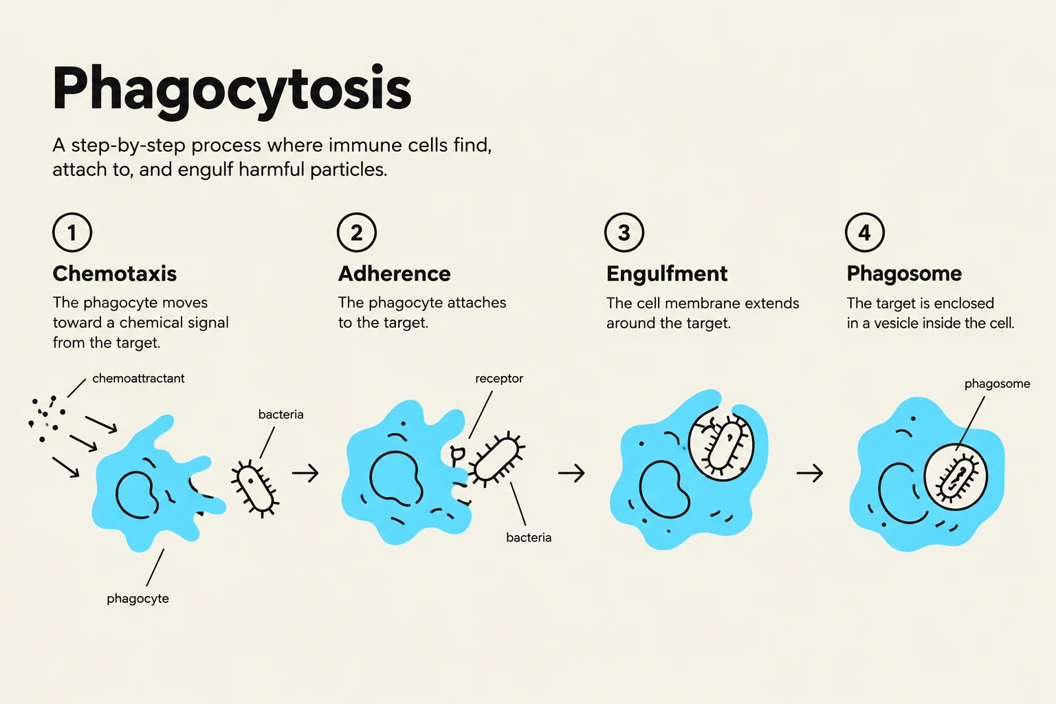
Phagocytes, such as macrophages (which can be "fixed" in tissues or "wandering" through them), destroy microbes through a process called phagocytosis. This process involves several critical stages:
Chemotaxis: Chemical signals attract phagocytes to the site of the microbe.
Adherence: The phagocyte attaches to the surface of the microorganism.
Ingestion: The microbe is taken into the cell. This can be aided by opsonization, where serum proteins coat the microbe to make it "tastier" for the phagocyte.
Digestion: The microbe is broken down inside a structure called a phagolysosome.
Phagocytosis is a step-by-step process where immune cells find, attach to, and engulf harmful particles. The diagram illustrates four stages: Chemotaxis, Adherence, Engulfment, and Phagosome.
When tissue is damaged or microbes are detected, the body initiates inflammation. Inflammation is characterized by four classic signs: redness (erythema), swelling (edema), pain, and heat. This process is driven by vasoactive mediators like histamine, kinins, prostaglandins, and leukotrienes, which increase blood vessel permeability to allow more immune cells to reach the site of infection.
Specialized Antimicrobial Mechanisms
Beyond cellular action, the body utilizes chemical and protein-based systems to fight infection.
The Complement System
The complement system consists of over 30 serum proteins produced by the liver that act in a "cascade"—one protein activates the next. This system assists in three major ways:
Cytolysis: Creating a "membrane attack complex" (MAC) that punches holes in microbial membranes, causing them to burst.
Opsonization: Coating microbes to enhance phagocytosis.
Inflammation: Activating mast cells to release histamine.
Interferons and Iron-Binding Proteins
To combat viruses, cells produce interferons (IFNs). Specifically, α and β interferons are produced in response to viral infections and signal neighboring cells to produce antiviral proteins (AVPs), which inhibit viral replication.
Finally, the body limits microbial growth by sequestering essential nutrients. Iron-binding proteins—such as transferrin (in blood), lactoferrin (in milk/saliva), and ferritin (in the liver/spleen)—bind to iron so tightly that bacteria cannot easily access it. Many bacteria attempt to counter this by producing siderophores to "steal" the iron back.
Diagram illustrating the three outcomes of complement activation: opsonization, inflammation, and lysis of a microbe.
Keep reading for free.
Create an account to finish this reading and make it part of your study routine.