Inflammatory Joint Diseases and Rheumatoid Arthritis
An overview of inflammatory joint diseases, focusing on Rheumatoid Arthritis (RA) and comparing it with other conditions like Ankylosing Spondylitis and Gout. Covers joint anatomy, RA pathogenesis, diagnosis, clinical manifestations, and extra-articular effects.
When we think of joint pain, we often imagine the "wear and tear" associated with aging. However, there is a much more aggressive category of disorders known as inflammatory joint diseases. Unlike simple mechanical degradation, these conditions are characterized by prominent inflammatory damage or destruction of the synovial membrane or articular cartilage, often accompanied by systemic signs of inflammation such as fever, malaise, and leukocytosis. Understanding the distinction between these systemic, immune-mediated processes and localized degenerative changes is crucial for clinical diagnosis and treatment.
Understanding the Joint Environment
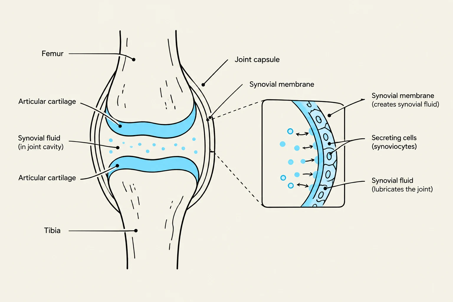
To understand how inflammation destroys a joint, we must first look at the healthy anatomy of a synovial joint (diarthrosis). In a healthy state, the articulating surfaces of bones are covered with avascular articular cartilage, which relies on the surrounding synovial fluid for oxygen and nutrients.
The synovial membrane is the inner layer of the joint capsule that secretes this vital synovial fluid. This fluid serves three primary purposes: nourishing the cartilage, reducing friction during movement, and absorbing shock. When this delicate balance is disrupted by an inflammatory process, the very structures meant to protect the joint become the site of its destruction.
Diagram illustrating the anatomy of a synovial joint, including the femur, tibia, articular cartilage, joint capsule, and synovial membrane with secreting cells (synoviocytes) that produce synovial fluid for lubrication.
Rheumatoid Arthritis (RA)
Rheumatoid arthritis is a chronic, multi-system inflammatory disease of unknown etiology, though it is widely considered a systemic autoimmune disorder. It primarily affects the synovial membrane, leading to persistent inflammatory synovitis.
Pathogenesis and the Pannus
The pathogenesis of RA involves a complex immune response. It is believed that a genetically susceptible individual is exposed to an arthritogenic antigen, causing a breakdown in self-tolerance. This triggers T-cells to accumulate in the joint, where they become sensitized to collagens, leading to a massive release of cytokines and the recruitment of further immune cells.
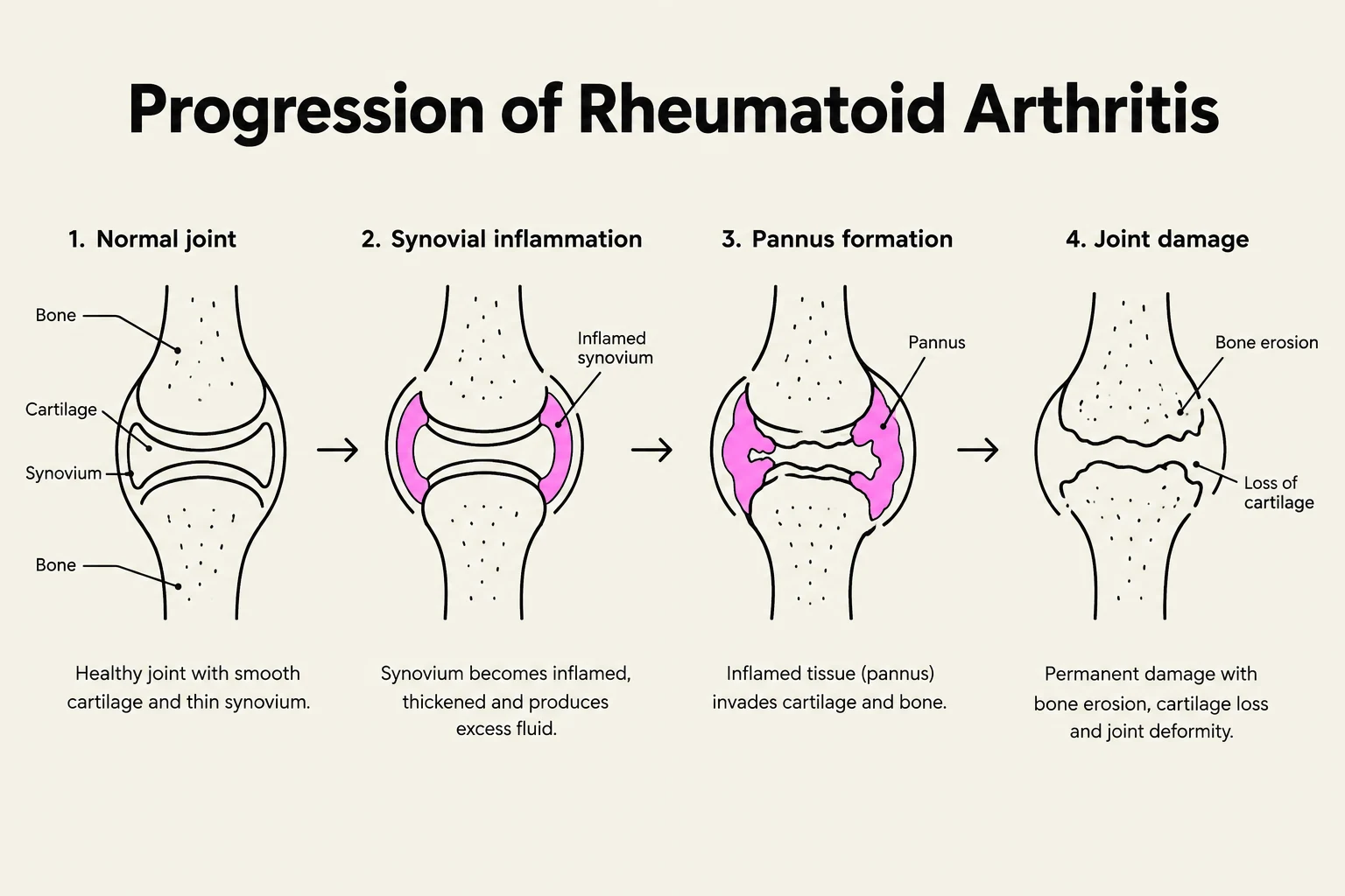
A hallmark of RA is the formation of a pannus. The pannus is an abnormal, thickened layer of fibrous tissue composed of inflammatory cells that release enzymes, oxidants, and cytokines. This aggressive tissue invades the joint space, causing the progressive loss of both bone and articular cartilage.
Auto-antibodies and Diagnosis
Clinicians often look for specific biomarkers to distinguish RA from other forms of arthritis. Approximately 80% of patients test positive for rheumatoid factor (RF), which is an auto-antibody directed against the Fc portion of IgG.
Exam Tip: To diagnose RA, clinicians often look for a cluster of symptoms lasting at least 6 weeks, including morning stiffness lasting over an hour, swelling in three or more joints (especially the hands), and the presence of rheumatoid nodules.
Clinical Manifestations
RA is characterized by polyarthritis—inflammation affecting multiple joints—which is typically symmetrical and affects peripheral joints. As the disease progresses, patients may develop significant joint deformities. Beyond the joints, RA can cause extra-articular manifestations, meaning the inflammation affects other organ systems. These can include:
Cardiovascular/Pulmonary: Pericarditis or pleural effusion.
Ocular: Scleritis.
Systemic: Fatigue, weight loss, and fever.
Renal: Nephropathy.
Diagram illustrating the progression of rheumatoid arthritis, showing stages from a normal joint to joint damage with bone erosion and cartilage loss.
Other Inflammatory Joint Diseases
While RA is a major driver of inflammatory joint disease, other conditions present with distinct pathological profiles.
Ankylosing Spondylitis
Ankylosing spondylitis is a chronic inflammatory disease specifically targeting the joints of the axial skeleton (the spine). It often begins in the sacroiliac joints and moves up the spinal column. The pathogenesis involves inflammation of the fibrocartilage, which leads to immune cell infiltration and cartilage erosion. As the body attempts to repair this damage, scar tissue calcifies, eventually causing the spinal column to fuse—a condition often referred to as a "bamboo spine".
Systemic Lupus Erythematosus (SLE)
Though often discussed alongside RA, Systemic Lupus Erythematosus is another example of an inflammatory disease that can manifest with joint involvement. Like RA, it is characterized by systemic inflammation, but its clinical presentation can vary widely across different organ systems.
Crystal-Induced Arthritis: Gout
Gout is a different type of inflammatory process driven by metabolic waste rather than direct autoimmunity. It is caused by the deposition of uric acid crystals within the synovial fluid. When these crystals are present, they trigger a massive white blood cell response, leading to intense, often sudden, joint inflammation.
Summary Comparison of Joint Diseases
The following table summarizes the key differences between the primary inflammatory and degenerative conditions discussed in the text.
Feature
Osteoarthritis (OA)
Rheumatoid Arthritis (RA)
Ankylosing Spondylitis
Gout
Primary Mechanism
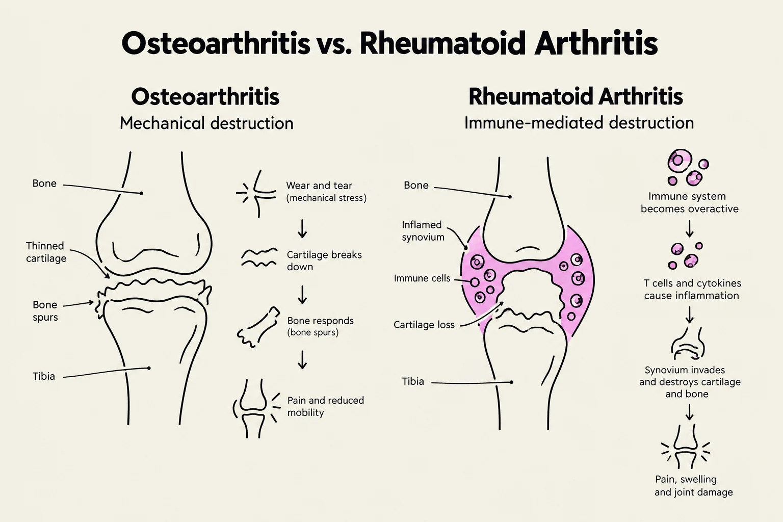
Mechanical "wear and tear" / Degenerative
Autoimmune / Pannus formation
Axial inflammation / Spinal fusion
Uric acid crystal deposition
Inflammation Type
Localized/Secondary
Systemic/Polyarthritis
Axial/Spinal
Acute/Crystal-induced
Key Biomarkers
Normal RF and ANA
Positive Rheumatoid Factor (RF)
HLA-B27 association
Elevated Uric Acid
Comparison of Osteoarthritis and Rheumatoid Arthritis, illustrating their distinct mechanisms of joint destruction.
Keep reading for free.
Create an account to finish this reading and make it part of your study routine.